 Movie
Movie Controller


+ Open data

- Basic information
| Entry | Database: PDB / ID: 2jc2 | ||||||
|---|---|---|---|---|---|---|---|
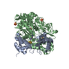
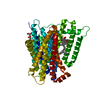
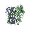
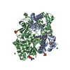
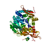
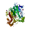
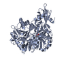
| Title | The crystal structure of the natural F112L human sorcin mutant | ||||||
 Components Components | SORCIN SRI (gene) SRI (gene) | ||||||
 Keywords Keywords | METAL BINDING PROTEIN / RYANODINE RECEPTOR INTERACTING PROTEIN / CALCIUM BINDING PROTEIN / NATURAL F112L SORCIN MUTANT / CALCIUM | ||||||
| Function / homology |  Function and homology information Function and homology informationregulation of relaxation of muscle / regulation of high voltage-gated calcium channel activity / regulation of cell communication by electrical coupling / regulation of striated muscle contraction / negative regulation of cardiac muscle contraction / regulation of cardiac muscle cell contraction / Sodium/Calcium exchangers / regulation of heart contraction / muscle organ development / Reduction of cytosolic Ca++ levels ...regulation of relaxation of muscle / regulation of high voltage-gated calcium channel activity / regulation of cell communication by electrical coupling / regulation of striated muscle contraction / negative regulation of cardiac muscle contraction / regulation of cardiac muscle cell contraction / Sodium/Calcium exchangers / regulation of heart contraction / muscle organ development / Reduction of cytosolic Ca++ levels / action potential / negative regulation of heart rate / regulation of cell communication by electrical coupling involved in cardiac conduction / positive regulation of insulin secretion involved in cellular response to glucose stimulus / regulation of calcium ion transport / Ion transport by P-type ATPases / intracellular sequestering of iron ion / calcium channel regulator activity / negative regulation of ryanodine-sensitive calcium-release channel activity / regulation of release of sequestered calcium ion into cytosol by sarcoplasmic reticulum / Ion homeostasis / T-tubule / sarcoplasmic reticulum membrane / positive regulation of release of sequestered calcium ion into cytosol / sarcoplasmic reticulum / Stimuli-sensing channels / Z disc / calcium ion transport / heart development / DNA-binding transcription factor binding / protease binding / transmembrane transporter binding / protein heterodimerization activity / signaling receptor binding / calcium ion binding / endoplasmic reticulum membrane / signal transduction / extracellular exosome / nucleoplasm / membrane / identical protein binding / cytosol / cytoplasmSimilarity search - Function | ||||||
| Biological species |  HOMO SAPIENS (human) HOMO SAPIENS (human) | ||||||
| Method | X-RAY DIFFRACTION / SYNCHROTRON / MOLECULAR REPLACEMENT / Resolution: 2.5 Å | ||||||
 Authors Authors | Franceschini, S. / Ilari, A. / Colotti, G. / Chiancone, E. | ||||||
 Citation Citation | Journal: Faseb J. / Year: 2008 Title: Molecular Basis for the Impaired Function of the Natural F112L Sorcin Mutant: X-Ray Crystal Structure, Calcium Affinity, and Interaction with Annexin Vii and the Ryanodine Receptor. Authors: Franceschini, S. / Ilari, A. / Verzili, D. / Zamparelli, C. / Antaramian, A. / Rueda, A. / Valdivia, H.H. / Chiancone, E. / Colotti, G. #1: Journal: J.Mol.Biol. / Year: 2002Title: The Crystal Structure of the Sorcin Calcium Binding Domain Provides a Model of Calcium-Dependent Processes in the Full Length Protein Authors: Ilari, A. / Johnson, K.A. / Nastopoulos, V. / Verzili, D. / Zamparelli, C. / Colotti, G. / Tsernoglou, D. / Chiancone, E. #2: Journal: Protein Sci. / Year: 2001Title: Crystal Structure of Calcium-Free Human Sorcin: A Member of the Penta-EF-Hand Protein Family Authors: Xie, X. / Dwyer, M.D. / Swenson, L. / Parker, M.H. / Botfield, M.C. | ||||||
| History |
|
- Structure visualization
| Structure viewer | Molecule: MolmilJmol/JSmol |
|---|
- Downloads & links
-Download
| PDBx/mmCIF format | 2jc2.cif.gz | 140.4 KB | Display | PDBx/mmCIF format |
|---|---|---|---|---|
| PDB format | pdb2jc2.ent.gz | 111.5 KB | Display | PDB format |
| PDBx/mmJSON format | 2jc2.json.gz | Tree view | PDBx/mmJSON format | |
| Others |  Other downloads Other downloads |
-Validation report
| Arichive directory | https://data.pdbj.org/pub/pdb/validation_reports/jc/2jc2ftp://data.pdbj.org/pub/pdb/validation_reports/jc/2jc2 | HTTPS FTP |
|---|
-Related structure data
| Related structure data |  1juoS S: Starting model for refinement |
|---|---|
| Similar structure data |


-Links
 PDBj
PDBj


- Assembly
| Deposited unit | 
| ||||||||||||||||||||||||||||||||||||||
|---|---|---|---|---|---|---|---|---|---|---|---|---|---|---|---|---|---|---|---|---|---|---|---|---|---|---|---|---|---|---|---|---|---|---|---|---|---|---|---|
| 1 | 
| ||||||||||||||||||||||||||||||||||||||
| 2 | 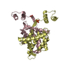
| ||||||||||||||||||||||||||||||||||||||
| Unit cell |
| ||||||||||||||||||||||||||||||||||||||
| Noncrystallographic symmetry (NCS) | NCS domain:
NCS domain segments: Component-ID: 1 / Beg auth comp-ID: ASP / Beg label comp-ID: ASP / End auth comp-ID: VAL / End label comp-ID: VAL / Refine code: 1 / Auth seq-ID: 33 - 198 / Label seq-ID: 33 - 198
NCS ensembles :
NCS oper: (Code: given Matrix: (0.50028, 0.86586, -0.00026), Vector : |
-Components
| #1: Protein | SRI (gene) / 22 KDA PROTEIN / CP-22 / V19 / F112L HUMAN SORCIN MUTANT Mass: 21661.320 Da / Num. of mol.: 4 / Mutation: YES Source method: isolated from a genetically manipulated source Source: (gene. exp.) HOMO SAPIENS (human) / Organ: HEART, MUSCLE, BRAIN AND ADRENAL MEDULLA / Plasmid: PET22B / Production host:  ESCHERICHIA COLI (E. coli) / Strain (production host): BL21(DE3) / References: UniProt: P30626 ESCHERICHIA COLI (E. coli) / Strain (production host): BL21(DE3) / References: UniProt: P30626#2: Chemical | ChemComp-SO4 / Sulfate  Mass: 96.063 Da / Num. of mol.: 6 / Source method: obtained synthetically / Formula: SO4 Mass: 96.063 Da / Num. of mol.: 6 / Source method: obtained synthetically / Formula: SO4#3: Water | ChemComp-HOH / | Water Mass: 18.015 Da / Num. of mol.: 140 / Source method: isolated from a natural source / Formula: H2O Mass: 18.015 Da / Num. of mol.: 140 / Source method: isolated from a natural source / Formula: H2OCompound details | ENGINEERED RESIDUE IN CHAIN A, PHE 112 TO LEU ENGINEERED RESIDUE IN CHAIN B, PHE 112 TO LEU ...ENGINEERED | Sequence details | F112L IS A NATURAL HUMAN SORCIN MUTANT. | |
|---|
-Experimental details
-Experiment
| Experiment | Method: X-RAY DIFFRACTION / Number of used crystals: 1 |
|---|
- Sample preparation
| Crystal | Density Matthews: 2.51 Å3/Da / Density % sol: 50.52 % / Description: NONE |
|---|---|
| Crystal grow | pH: 5.7 / Details: AMMONIUM SULFATE 1.0M, PH 5.7, 12% V/V DIOXANE |
-Data collection
| Diffraction | Mean temperature: 100 K |
|---|---|
| Diffraction source | Source: SYNCHROTRON / Site: ESRF  / Beamline: ID14-2 / Wavelength: 0.934 / Beamline: ID14-2 / Wavelength: 0.934 |
| Detector | Type: ADSC CCD / Detector: CCD |
| Radiation | Protocol: SINGLE WAVELENGTH / Monochromatic (M) / Laue (L): M / Scattering type: x-ray |
| Radiation wavelength | Wavelength: 0.934 Å / Relative weight: 1 |
| Reflection | Resolution: 2.5→50 Å / Num. obs: 25105 / % possible obs: 96.8 % / Redundancy: 5.2 % / Rmerge(I) obs: 0.08 / Net I/σ(I): 7 |
| Reflection shell | Resolution: 2.5→2.59 Å / Redundancy: 3.3 % / Rmerge(I) obs: 0.6 / Mean I/σ(I) obs: 2 / % possible all: 85.1 |
- Processing
| Software |
| ||||||||||||||||||||||||||||||||||||||||||||||||||||||||||||||||||||||||||||||||||||||||||||||||||||||||||||||||||||||||||||||||||||||||||||||||||||||||||||||||||||||||||||||||||||||
|---|---|---|---|---|---|---|---|---|---|---|---|---|---|---|---|---|---|---|---|---|---|---|---|---|---|---|---|---|---|---|---|---|---|---|---|---|---|---|---|---|---|---|---|---|---|---|---|---|---|---|---|---|---|---|---|---|---|---|---|---|---|---|---|---|---|---|---|---|---|---|---|---|---|---|---|---|---|---|---|---|---|---|---|---|---|---|---|---|---|---|---|---|---|---|---|---|---|---|---|---|---|---|---|---|---|---|---|---|---|---|---|---|---|---|---|---|---|---|---|---|---|---|---|---|---|---|---|---|---|---|---|---|---|---|---|---|---|---|---|---|---|---|---|---|---|---|---|---|---|---|---|---|---|---|---|---|---|---|---|---|---|---|---|---|---|---|---|---|---|---|---|---|---|---|---|---|---|---|---|---|---|---|---|
| Refinement | Method to determine structure: MOLECULAR REPLACEMENT Starting model: PDB ENTRY 1JUO Resolution: 2.5→45.5 Å / Cor.coef. Fo:Fc: 0.909 / Cor.coef. Fo:Fc free: 0.887 / SU B: 12.596 / SU ML: 0.287 / Cross valid method: THROUGHOUT / ESU R: 1.059 / ESU R Free: 0.355 / Stereochemistry target values: MAXIMUM LIKELIHOOD Details: HYDROGENS HAVE BEEN ADDED IN THE RIDING POSITIONS. RESIDUES FROM M1 TO Q32 ARE MISSING IN THE STRUCTURE BECAUSE OF AN INSUFFICIENT ELECTRON DENSITY
| ||||||||||||||||||||||||||||||||||||||||||||||||||||||||||||||||||||||||||||||||||||||||||||||||||||||||||||||||||||||||||||||||||||||||||||||||||||||||||||||||||||||||||||||||||||||
| Solvent computation | Ion probe radii: 0.8 Å / Shrinkage radii: 0.8 Å / VDW probe radii: 1.4 Å / Solvent model: MASK | ||||||||||||||||||||||||||||||||||||||||||||||||||||||||||||||||||||||||||||||||||||||||||||||||||||||||||||||||||||||||||||||||||||||||||||||||||||||||||||||||||||||||||||||||||||||
| Displacement parameters | Biso mean: 38.97 Å2
| ||||||||||||||||||||||||||||||||||||||||||||||||||||||||||||||||||||||||||||||||||||||||||||||||||||||||||||||||||||||||||||||||||||||||||||||||||||||||||||||||||||||||||||||||||||||
| Refinement step | Cycle: LAST / Resolution: 2.5→45.5 Å
| ||||||||||||||||||||||||||||||||||||||||||||||||||||||||||||||||||||||||||||||||||||||||||||||||||||||||||||||||||||||||||||||||||||||||||||||||||||||||||||||||||||||||||||||||||||||
| Refine LS restraints |
|