 Movie
Movie Controller

[English] 日本語
 Yorodumi
- PDB-2j4a: Human Thyroid hormone receptor beta ligand binding domain in comp... -
+ Open data

- Basic information
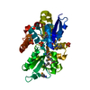
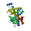
| Entry | Database: PDB / ID: 2j4a | ||||||
|---|---|---|---|---|---|---|---|
| Title | Human Thyroid hormone receptor beta ligand binding domain in complex with KB131084 | ||||||
 Components Components | THYROID HORMONE RECEPTOR BETA-1 | ||||||
 Keywords Keywords | RECEPTOR / NUCLEAR PROTEIN / DISEASE MUTATION / NUCLEAR RECEPTOR / ZINC / DEAFNESS / ZINC-FINGER / DNA-BINDING / TRANSCRIPTION REGULATION / TRANSCRIPTION / ALTERNATIVE SPLICING / LIGAND BINDING DOMAIN / POLYMORPHISM / METAL-BINDING | ||||||
| Function / homology |  Function and homology information Function and homology informationretinal cone cell apoptotic process / negative regulation of female receptivity / female courtship behavior / retinal cone cell development / thyroid hormone mediated signaling pathway / positive regulation of thyroid hormone mediated signaling pathway / cellular response to thyroid hormone stimulus / regulation of heart contraction / type I pneumocyte differentiation / thyroid hormone binding ...retinal cone cell apoptotic process / negative regulation of female receptivity / female courtship behavior / retinal cone cell development / thyroid hormone mediated signaling pathway / positive regulation of thyroid hormone mediated signaling pathway / cellular response to thyroid hormone stimulus / regulation of heart contraction / type I pneumocyte differentiation / thyroid hormone binding / retinoic acid receptor signaling pathway / sensory perception of sound / SUMOylation of intracellular receptors / mRNA transcription by RNA polymerase II / transcription coactivator binding / chromatin DNA binding / Nuclear Receptor transcription pathway / RNA polymerase II transcription regulator complex / nuclear receptor activity / sequence-specific double-stranded DNA binding / cell differentiation / nuclear body / DNA-binding transcription factor activity, RNA polymerase II-specific / RNA polymerase II cis-regulatory region sequence-specific DNA binding / DNA-binding transcription factor activity / DNA-templated transcription / chromatin / negative regulation of transcription by RNA polymerase II / enzyme binding / positive regulation of transcription by RNA polymerase II / DNA binding / zinc ion binding / nucleoplasm / nucleusSimilarity search - Function | ||||||
| Biological species |  HOMO SAPIENS (human) HOMO SAPIENS (human) | ||||||
| Method | X-RAY DIFFRACTION / SYNCHROTRON / OTHER / Resolution: 2.2 Å | ||||||
 Authors Authors | Farnegardh, M. | ||||||
 Citation Citation | Journal: J.Med.Chem. / Year: 2006 Title: Thyroid Receptor Ligands. 6. A High Affinity "Direct Antagonist" Selective for the Thyroid Hormone Receptor. Authors: Koehler, K. / Gordon, S. / Brandt, P. / Carlsson, B. / Backsbro-Saedi, A. / Apelqvist, T. / Agback, P. / Grover, G.J. / Nelson, W. / Grynfarb, M. / Farnegardh, M. / Rehnmark, S. / Malm, J. | ||||||
| History |
|
- Structure visualization
| Structure viewer | Molecule: MolmilJmol/JSmol |
|---|
- Downloads & links
-Download
| PDBx/mmCIF format | 2j4a.cif.gz | 64.5 KB | Display | PDBx/mmCIF format |
|---|---|---|---|---|
| PDB format | pdb2j4a.ent.gz | 47 KB | Display | PDB format |
| PDBx/mmJSON format | 2j4a.json.gz | Tree view | PDBx/mmJSON format | |
| Others |  Other downloads Other downloads |
-Validation report
| Arichive directory | https://data.pdbj.org/pub/pdb/validation_reports/j4/2j4aftp://data.pdbj.org/pub/pdb/validation_reports/j4/2j4a | HTTPS FTP |
|---|
-Related structure data
| Related structure data | |
|---|---|
| Similar structure data |


-Links
 PDBj
PDBj
- Assembly
| Deposited unit | 
| ||||||||
|---|---|---|---|---|---|---|---|---|---|
| 1 |
| ||||||||
| Unit cell |
| ||||||||
| Components on special symmetry positions |
|
-Components
| #1: Protein | / THYROID HORMONE RECEPTOR BETA LBD Mass: 28555.057 Da / Num. of mol.: 1 / Fragment: LIGAND BINDING DOMAIN, RESIDUES 209-461 / Mutation: YES Source method: isolated from a genetically manipulated source Source: (gene. exp.) HOMO SAPIENS (human) / Production host:  ESCHERICHIA COLI (E. coli) / References: UniProt: P10828 ESCHERICHIA COLI (E. coli) / References: UniProt: P10828 |
|---|---|
| #2: Chemical | ChemComp-OEF / 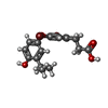  Mass: 458.141 Da / Num. of mol.: 1 / Source method: obtained synthetically / Formula: C18H18Br2O4 Mass: 458.141 Da / Num. of mol.: 1 / Source method: obtained synthetically / Formula: C18H18Br2O4 |
| #3: Water | ChemComp-HOH / Water Mass: 18.015 Da / Num. of mol.: 107 / Source method: isolated from a natural source / Formula: H2O Mass: 18.015 Da / Num. of mol.: 107 / Source method: isolated from a natural source / Formula: H2O |
| Compound details | ENGINEERED |
-Experimental details
-Experiment
| Experiment | Method: X-RAY DIFFRACTION |
|---|
- Sample preparation
| Crystal | Density Matthews: 2.37 Å3/Da / Density % sol: 47.8 % / Description: NONE |
|---|---|
| Crystal grow | Details: 0.45 M AMMONIUMSULFATE, 100 MMTRIS PH8.0, 100MM NDSB |
-Data collection
| Diffraction | Mean temperature: 100 K |
|---|---|
| Diffraction source | Source: SYNCHROTRON / Site: EMBL/DESY, HAMBURG  / Beamline: BW7A / Wavelength: 0.933 / Beamline: BW7A / Wavelength: 0.933 |
| Radiation | Protocol: SINGLE WAVELENGTH / Monochromatic (M) / Laue (L): M / Scattering type: x-ray |
| Radiation wavelength | Wavelength: 0.933 Å / Relative weight: 1 |
| Reflection | Resolution: 2.2→20 Å / Num. obs: 14513 / % possible obs: 99.9 % / Observed criterion σ(I): 0 / Redundancy: 5.1 % / Biso Wilson estimate: 20.4 Å2 / Rmerge(I) obs: 0.11 / Net I/σ(I): 7.7 |
- Processing
| Software | Name: CNS / Classification: refinement | ||||||||||||||||||||||||||||||||||||||||||||||||||||||||||||||||||||||||||||||||
|---|---|---|---|---|---|---|---|---|---|---|---|---|---|---|---|---|---|---|---|---|---|---|---|---|---|---|---|---|---|---|---|---|---|---|---|---|---|---|---|---|---|---|---|---|---|---|---|---|---|---|---|---|---|---|---|---|---|---|---|---|---|---|---|---|---|---|---|---|---|---|---|---|---|---|---|---|---|---|---|---|---|
| Refinement | Method to determine structure: OTHER / Resolution: 2.2→40 Å / Rfactor Rfree error: 0.01 / Data cutoff high absF: 10000 / Isotropic thermal model: RESTRAINED / Cross valid method: THROUGHOUT / σ(F): 0 / Details: BULK SOLVENT MODEL USED
| ||||||||||||||||||||||||||||||||||||||||||||||||||||||||||||||||||||||||||||||||
| Solvent computation | Solvent model: FLAT MODEL / Bsol: 35.8737 Å2 / ksol: 0.343697 e/Å3 | ||||||||||||||||||||||||||||||||||||||||||||||||||||||||||||||||||||||||||||||||
| Displacement parameters | Biso mean: 31.9 Å2
| ||||||||||||||||||||||||||||||||||||||||||||||||||||||||||||||||||||||||||||||||
| Refine analyze |
| ||||||||||||||||||||||||||||||||||||||||||||||||||||||||||||||||||||||||||||||||
| Refinement step | Cycle: LAST / Resolution: 2.2→40 Å
| ||||||||||||||||||||||||||||||||||||||||||||||||||||||||||||||||||||||||||||||||
| Refine LS restraints |
| ||||||||||||||||||||||||||||||||||||||||||||||||||||||||||||||||||||||||||||||||
| LS refinement shell | Resolution: 2.2→2.34 Å / Rfactor Rfree error: 0.032 / Total num. of bins used: 6
| ||||||||||||||||||||||||||||||||||||||||||||||||||||||||||||||||||||||||||||||||
| Xplor file |
|
