 Movie
Movie Controller

[English] 日本語
 Yorodumi
- PDB-4irm: Crystal structure of mntc r116a mutant exhibits flexibility in th... -
+ Open data

- Basic information
| Entry | Database: PDB / ID: 4irm | |||||||||
|---|---|---|---|---|---|---|---|---|---|---|
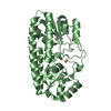
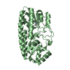
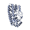
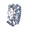
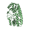
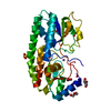
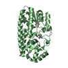
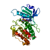
| Title | Crystal structure of mntc r116a mutant exhibits flexibility in the c-terminal domain | |||||||||
 Components Components | Mn transporter; MntC | |||||||||
 Keywords Keywords |  TRANSPORT PROTEIN / manganese / solute binding protein of ABS transporter TRANSPORT PROTEIN / manganese / solute binding protein of ABS transporter | |||||||||
| Function / homology |  Function and homology information Function and homology information | |||||||||
| Biological species |  | |||||||||
| Method | X-RAY DIFFRACTION / SYNCHROTRON / MOLECULAR REPLACEMENT / Resolution: 3.5 Å | |||||||||
 Authors Authors | Kanteev, M. / Adir, N. | |||||||||
 Citation Citation | Journal: Acta Crystallogr.,Sect.F / Year: 2013 Title: Arginine 116 stabilizes the entrance to the metal ion-binding site of the MntC protein. Authors: Kanteev, M. / Adir, N. | |||||||||
| History |
|
- Structure visualization
| Structure viewer | Molecule: MolmilJmol/JSmol |
|---|
- Downloads & links
-Download
| PDBx/mmCIF format | 4irm.cif.gz | 160.6 KB | Display | PDBx/mmCIF format |
|---|---|---|---|---|
| PDB format | pdb4irm.ent.gz | 125.5 KB | Display | PDB format |
| PDBx/mmJSON format | 4irm.json.gz | Tree view | PDBx/mmJSON format | |
| Others |  Other downloads Other downloads |
-Validation report
| Arichive directory | https://data.pdbj.org/pub/pdb/validation_reports/ir/4irmftp://data.pdbj.org/pub/pdb/validation_reports/ir/4irm | HTTPS FTP |
|---|
-Related structure data
| Related structure data |  3ujpSC S: Starting model for refinement C: citing same article ( |
|---|---|
| Similar structure data |


-Links
 PDBj
PDBj- Assembly
| Deposited unit | 
| ||||||||
|---|---|---|---|---|---|---|---|---|---|
| 1 | 
| ||||||||
| 2 | 
| ||||||||
| 3 | 
| ||||||||
| Unit cell |
|
-Components
| #1: Protein | Mass: 36014.133 Da / Num. of mol.: 3 / Mutation: R116A Source method: isolated from a genetically manipulated source Details: includes TEV cleavage site / Source: (gene. exp.) Escherichia coli (E. coli) / Strain (production host): BL21 / References: UniProt: Q79EF9#2: Chemical |   Mass: 54.938 Da / Num. of mol.: 3 / Source method: obtained synthetically / Formula: Mn Mass: 54.938 Da / Num. of mol.: 3 / Source method: obtained synthetically / Formula: Mn |
|---|
-Experimental details
-Experiment
| Experiment | Method: X-RAY DIFFRACTION / Number of used crystals: 1 |
|---|
- Sample preparation
| Crystal | Density Matthews: 1.99 Å3/Da / Density % sol: 38.15 % |
|---|---|
| Crystal grow | Temperature: 290 K / pH: 6.5 Details: PEG 4000, SODIUM CACODYLATE, 50mM ZINC CHLORIDE,, pH 6.5, VAPOR DIFFUSION, HANGING DROP, temperature 290K |
-Data collection
| Diffraction | Mean temperature: 100 K |
|---|---|
| Diffraction source | Source: SYNCHROTRON / Site: ESRF  / Beamline: ID23-1 / Wavelength: 0.934 / Beamline: ID23-1 / Wavelength: 0.934 |
| Detector | Type: ADSC QUANTUM 315r / Detector: CCD |
| Radiation | Protocol: SINGLE WAVELENGTH / Monochromatic (M) / Laue (L): M / Scattering type: x-ray |
| Radiation wavelength | Wavelength: 0.934 Å / Relative weight: 1 |
| Reflection | Resolution: 3.5→24.5 Å / Num. obs: 11028 / % possible obs: 99 % / Redundancy: 5.3 % / Rmerge(I) obs: 0.078 / Net I/σ(I): 15 |
| Reflection shell | Resolution: 3.5→3.69 Å / Redundancy: 4.9 % / Mean I/σ(I) obs: 6.2 |
- Processing
| Software |
| |||||||||||||||||||||||||||||||||||
|---|---|---|---|---|---|---|---|---|---|---|---|---|---|---|---|---|---|---|---|---|---|---|---|---|---|---|---|---|---|---|---|---|---|---|---|---|
| Refinement | Method to determine structure: MOLECULAR REPLACEMENT Starting model: PDB ENTRY 3UJP Resolution: 3.5→24.5 Å / Isotropic thermal model: Isotropic / Phase error: 35.89 / Stereochemistry target values: TWIN_LSQ_F
| |||||||||||||||||||||||||||||||||||
| Solvent computation | Shrinkage radii: 0.86 Å / VDW probe radii: 1.1 Å / Solvent model: FLAT BULK SOLVENT MODEL / Bsol: 64.19 Å2 / ksol: 0.24 e/Å3 | |||||||||||||||||||||||||||||||||||
| Displacement parameters | Biso mean: 97.5 Å2 | |||||||||||||||||||||||||||||||||||
| Refinement step | Cycle: LAST / Resolution: 3.5→24.5 Å
| |||||||||||||||||||||||||||||||||||
| Refine LS restraints |
| |||||||||||||||||||||||||||||||||||
| LS refinement shell |
|
