flap endonuclease activity / 5'-flap endonuclease activity / DNA replication, removal of RNA primer / 5'-3' exonuclease activity / DNA polymerase processivity factor activity / leading strand elongation / regulation of DNA replication / mismatch repair / translesion synthesis / DNA replication ...flap endonuclease activity / 5'-flap endonuclease activity / DNA replication, removal of RNA primer / 5'-3' exonuclease activity / DNA polymerase processivity factor activity / leading strand elongation / regulation of DNA replication / mismatch repair / translesion synthesis / DNA replication / Hydrolases; Acting on ester bonds / DNA repair / magnesium ion binding / DNA binding Similarity search - Function
SHEET THE SHEET STRUCTURE OF THIS MOLECULE IS BIFURCATED. IN ORDER TO REPRESENT THIS FEATURE IN ... SHEET THE SHEET STRUCTURE OF THIS MOLECULE IS BIFURCATED. IN ORDER TO REPRESENT THIS FEATURE IN THE SHEET RECORDS BELOW, TWO SHEETS ARE DEFINED.
Mass: 18.015 Da / Num. of mol.: 78 / Source method: isolated from a natural source / Formula: H2O
-
Details
Compound details
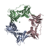
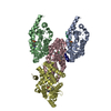
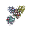
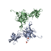
CHAIN A: ENDONUCLEASE THAT CLEAVES THE 5' OVERHANGING FLAP STRUCTURE THAT IS GENERATED BY ...CHAIN A: ENDONUCLEASE THAT CLEAVES THE 5' OVERHANGING FLAP STRUCTURE THAT IS GENERATED BY DISPLACEMENT SYNTHESIS WHEN DNA POLYMERASE ENCOUNTERS THE 5' END OF A DOWNSTREAM OKAZAKI FRAGMENT. CHAIN B: SLIDING CLAMP SUBUNIT. CHAIN C: SLIDING CLAMP SUBUNIT. RESPONSIBLE FOR TETHERING THE CATALYTIC SUBUNIT OF DNA POLYMERASE TO DNA DURING HIGH-SPEED REPLICATION.
Sequence details
FIRST FOUR RESIDUES REMOVED FOR CONSTRUCT
-
Experimental details
-
Experiment
Experiment
Method: X-RAY DIFFRACTION / Number of used crystals: 1
-
Sample preparation
Crystal
Density Matthews: 2.45 Å3/Da / Density % sol: 50 %
Crystal grow
Method: vapor diffusion, hanging drop Details: PRIOR TO CRYSTALLISATION THE PCNA1-PCNA2-PCNA3 COMPLEX WAS MIXED WITH FEN1 AT AN EQUIMOLAR RATIO AND INCUBATED FOR 20 MIN AT 294 K. PCNA1-PCNA2-FEN1 CO-CRYSTALS WERE GROWN BY THE VAPOUR ...Details: PRIOR TO CRYSTALLISATION THE PCNA1-PCNA2-PCNA3 COMPLEX WAS MIXED WITH FEN1 AT AN EQUIMOLAR RATIO AND INCUBATED FOR 20 MIN AT 294 K. PCNA1-PCNA2-FEN1 CO-CRYSTALS WERE GROWN BY THE VAPOUR DIFFUSION METHOD IN HANGING DROPS. PCNA3 WAS NOT PRESENT IN THE CRYSTAL LATTICE. DROPS WERE PREPARED BY MIXING 100 MM PROTEIN COMPLEX IN 20 MM TRIS-HCL PH 8.0, 200 MM NACL, 5 MM MGCL2, 2 MM DTT BUFFER SOLUTION WITH EQUAL VOLUMES OF 0.1M ACETATE PH 4.8, 8% PEG 8,000, 220 MM ZNOAC2 AND 30 MM GLYCYL-GLYCYL-GLYCINE.
 Movie
Movie Controller


 Open data
 Basic information
 Components
 Keywords
 HYDROLASE /
  Function and homology information

 Authors
 Citation
 Structure visualization
 Downloads & links
 Other downloads
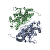


 PDBj
PDBj


 Assembly


 Mass: 65.409 Da / Num. of mol.: 4 / Source method: obtained synthetically / Formula: Zn
 Mass: 24.305 Da / Num. of mol.: 4 / Source method: obtained synthetically / Formula: Mg
 Sample preparation
 / Beamline: ID29 / Wavelength: 0.9795
  Processing