 Movie
Movie Controller


+ Open data

- Basic information
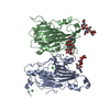
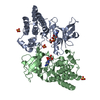
| Entry | Database: PDB / ID: 2ivt | ||||||
|---|---|---|---|---|---|---|---|
| Title | Crystal structure of phosphorylated RET tyrosine kinase domain | ||||||
 Components Components | PROTO-ONCOGENE TYROSINE-PROTEIN KINASE RECEPTOR RET PRECURSOR | ||||||
 Keywords Keywords |  TRANSFERASE / NUCLEOTIDE-BINDING / HIRSCHSPRUNG DISEASE / PHOSPHORYLATION / DISEASE MUTATION / PHOSPHOTRANSFERASE / TYROSINE-PROTEIN KINASE / CHROMOSOMAL TRANSLOCATION / POLYMORPHISM / GDNF RECEPTOR / TRANSMEMBRANE / PROTO-ONCOGENE / TYROSINE KINASE / RET / KINASE / MEMBRANE / ATP-BINDING TRANSFERASE / NUCLEOTIDE-BINDING / HIRSCHSPRUNG DISEASE / PHOSPHORYLATION / DISEASE MUTATION / PHOSPHOTRANSFERASE / TYROSINE-PROTEIN KINASE / CHROMOSOMAL TRANSLOCATION / POLYMORPHISM / GDNF RECEPTOR / TRANSMEMBRANE / PROTO-ONCOGENE / TYROSINE KINASE / RET / KINASE / MEMBRANE / ATP-BINDING | ||||||
| Function / homology |  Function and homology information Function and homology informationglial cell-derived neurotrophic factor receptor signaling pathway / Peyer's patch morphogenesis / positive regulation of metanephric glomerulus development / posterior midgut development / ureter maturation / embryonic epithelial tube formation / lymphocyte migration into lymphoid organs / membrane protein proteolysis / positive regulation of peptidyl-serine phosphorylation of STAT protein / positive regulation of neuron maturation ...glial cell-derived neurotrophic factor receptor signaling pathway / Peyer's patch morphogenesis / positive regulation of metanephric glomerulus development / posterior midgut development / ureter maturation / embryonic epithelial tube formation / lymphocyte migration into lymphoid organs / membrane protein proteolysis / positive regulation of peptidyl-serine phosphorylation of STAT protein / positive regulation of neuron maturation / neuron cell-cell adhesion / enteric nervous system development / innervation / plasma membrane protein complex / neuron maturation / positive regulation of extrinsic apoptotic signaling pathway in absence of ligand / positive regulation of cell adhesion mediated by integrin / neural crest cell migration / ureteric bud development / response to pain / regulation of axonogenesis / homophilic cell adhesion via plasma membrane adhesion molecules / positive regulation of cell size / RET signaling / regulation of cell adhesion / cellular response to retinoic acid / NPAS4 regulates expression of target genes / transmembrane receptor protein tyrosine kinase activity / axon guidance / receptor protein-tyrosine kinase / positive regulation of neuron projection development / cell surface receptor protein tyrosine kinase signaling pathway / activation of cysteine-type endopeptidase activity involved in apoptotic process / MAPK cascade / retina development in camera-type eye / signaling receptor activity / RAF/MAP kinase cascade / protein tyrosine kinase activity / positive regulation of MAPK cascade / positive regulation of phosphatidylinositol 3-kinase/protein kinase B signal transduction / receptor complex / early endosome / endosome membrane / positive regulation of cell migration / response to xenobiotic stimulus / axon / protein phosphorylation / neuronal cell body / dendrite / calcium ion binding / positive regulation of gene expression / positive regulation of DNA-templated transcription / signal transduction / ATP binding / plasma membraneSimilarity search - Function | ||||||
| Biological species |  HOMO SAPIENS (human) HOMO SAPIENS (human) | ||||||
| Method | X-RAY DIFFRACTION / SYNCHROTRON / MOLECULAR REPLACEMENT / Resolution: 2.6 Å | ||||||
 Authors Authors | Knowles, P.P. / Murray-Rust, J. / McDonald, N.Q. | ||||||
 Citation Citation | Journal: J. Biol. Chem. / Year: 2006 Title: Structure and chemical inhibition of the RET tyrosine kinase domain. Authors: Knowles, P.P. / Murray-Rust, J. / Kjaer, S. / Scott, R.P. / Hanrahan, S. / Santoro, M. / Ibanez, C.F. / McDonald, N.Q. | ||||||
| History |
|
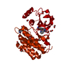
- Structure visualization
| Structure viewer | Molecule: MolmilJmol/JSmol |
|---|
- Downloads & links
-Download
| PDBx/mmCIF format | 2ivt.cif.gz | 74.4 KB | Display | PDBx/mmCIF format |
|---|---|---|---|---|
| PDB format | pdb2ivt.ent.gz | 52.6 KB | Display | PDB format |
| PDBx/mmJSON format | 2ivt.json.gz | Tree view | PDBx/mmJSON format | |
| Others |  Other downloads Other downloads |
-Validation report
| Arichive directory | https://data.pdbj.org/pub/pdb/validation_reports/iv/2ivtftp://data.pdbj.org/pub/pdb/validation_reports/iv/2ivt | HTTPS FTP |
|---|
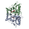
-Related structure data
| Related structure data |  2ivsC  2ivuC  2ivvC 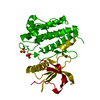 1gjoS C: citing same article ( S: Starting model for refinement |
|---|---|
| Similar structure data |


-Links
 PDBj
PDBj


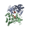
- Assembly
| Deposited unit | 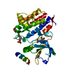
| ||||||||
|---|---|---|---|---|---|---|---|---|---|
| 1 | 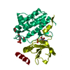
| ||||||||
| Unit cell |
|
-Components
| #1: Protein | Mass: 35788.215 Da / Num. of mol.: 1 / Fragment: TYROSINE KINASE DOMAIN, RESIDUES 705-1013 Source method: isolated from a genetically manipulated source Details: PTR AT 905 / Source: (gene. exp.) HOMO SAPIENS (human) / Plasmid: PBACPAK-HIS3 / Cell line (production host): SF9 / Production host:   SPODOPTERA FRUGIPERDA (fall armyworm) SPODOPTERA FRUGIPERDA (fall armyworm)References: UniProt: P07949, receptor protein-tyrosine kinase | ||||||
|---|---|---|---|---|---|---|---|
| #2: Chemical | Formic acid  Mass: 46.025 Da / Num. of mol.: 2 / Source method: obtained synthetically / Formula: CH2O2 Mass: 46.025 Da / Num. of mol.: 2 / Source method: obtained synthetically / Formula: CH2O2#3: Chemical | ChemComp-AMP / | Adenosine monophosphate  Mass: 347.221 Da / Num. of mol.: 1 / Source method: obtained synthetically / Formula: C10H14N5O7P / Comment: AMP*YM Mass: 347.221 Da / Num. of mol.: 1 / Source method: obtained synthetically / Formula: C10H14N5O7P / Comment: AMP*YM#4: Water | ChemComp-HOH / | Water Mass: 18.015 Da / Num. of mol.: 62 / Source method: isolated from a natural source / Formula: H2O Mass: 18.015 Da / Num. of mol.: 62 / Source method: isolated from a natural source / Formula: H2OSequence details | 705-1013 CORRESPOND | |
-Experimental details
-Experiment
| Experiment | Method: X-RAY DIFFRACTION / Number of used crystals: 1 |
|---|
- Sample preparation
| Crystal | Density Matthews: 2.76 Å3/Da / Density % sol: 57.7 % |
|---|---|
| Crystal grow | Temperature: 295 K / Method: vapor diffusion, sitting drop Details: PROTEIN 3 MG/ML IN 20 MM TRIS-HCL PH 8.0, 100MM NACL, 1MM DTT RESERVOIR 2.0 M SODIUM FORMATE, 0.1M SODIUM CITRATE PH 5.5,2.5MM ATP 5MM MAGNESIUM CHLORIDE VAPOUR DIFFUSION, SITTING DROP, 295 K |
-Data collection
| Diffraction | Mean temperature: 100 K |
|---|---|
| Diffraction source | Source: SYNCHROTRON / Site: SRS  / Beamline: PX14.2 / Wavelength: 0.978 / Beamline: PX14.2 / Wavelength: 0.978 |
| Detector | Type: ADSC CCD / Detector: CCD / Date: Jun 25, 2003 |
| Radiation | Protocol: SINGLE WAVELENGTH / Monochromatic (M) / Laue (L): M / Scattering type: x-ray |
| Radiation wavelength | Wavelength: 0.978 Å / Relative weight: 1 |
| Reflection | Resolution: 2.6→39.5 Å / Num. obs: 12129 / % possible obs: 100 % / Observed criterion σ(I): 0 / Redundancy: 3.6 % / Rmerge(I) obs: 0.05 / Net I/σ(I): 12.4 |
| Reflection shell | Resolution: 2.6→2.74 Å / Redundancy: 3.7 % / Rmerge(I) obs: 0.13 / Mean I/σ(I) obs: 5.1 / % possible all: 100 |
- Processing
| Software |
| ||||||||||||||||||||||||||||||||||||||||||||||||||||||||||||||||||||||||||||||||||||||||||||||||||||||||||||||||||||||||||||||||||||||||||||||||||||||||||||||||||||||||||||||||||||||
|---|---|---|---|---|---|---|---|---|---|---|---|---|---|---|---|---|---|---|---|---|---|---|---|---|---|---|---|---|---|---|---|---|---|---|---|---|---|---|---|---|---|---|---|---|---|---|---|---|---|---|---|---|---|---|---|---|---|---|---|---|---|---|---|---|---|---|---|---|---|---|---|---|---|---|---|---|---|---|---|---|---|---|---|---|---|---|---|---|---|---|---|---|---|---|---|---|---|---|---|---|---|---|---|---|---|---|---|---|---|---|---|---|---|---|---|---|---|---|---|---|---|---|---|---|---|---|---|---|---|---|---|---|---|---|---|---|---|---|---|---|---|---|---|---|---|---|---|---|---|---|---|---|---|---|---|---|---|---|---|---|---|---|---|---|---|---|---|---|---|---|---|---|---|---|---|---|---|---|---|---|---|---|---|
| Refinement | Method to determine structure: MOLECULAR REPLACEMENT Starting model: PDB ENTRY 1GJO Resolution: 2.6→50 Å / Cor.coef. Fo:Fc: 0.937 / Cor.coef. Fo:Fc free: 0.89 / SU B: 9.87 / SU ML: 0.213 / Cross valid method: THROUGHOUT / ESU R: 0.496 / ESU R Free: 0.295 / Stereochemistry target values: MAXIMUM LIKELIHOOD / Details: HYDROGENS HAVE BEEN ADDED IN THE RIDING POSITIONS.
| ||||||||||||||||||||||||||||||||||||||||||||||||||||||||||||||||||||||||||||||||||||||||||||||||||||||||||||||||||||||||||||||||||||||||||||||||||||||||||||||||||||||||||||||||||||||
| Solvent computation | Ion probe radii: 0.8 Å / Shrinkage radii: 0.8 Å / VDW probe radii: 1.2 Å / Solvent model: MASK | ||||||||||||||||||||||||||||||||||||||||||||||||||||||||||||||||||||||||||||||||||||||||||||||||||||||||||||||||||||||||||||||||||||||||||||||||||||||||||||||||||||||||||||||||||||||
| Displacement parameters | Biso mean: 23.25 Å2
| ||||||||||||||||||||||||||||||||||||||||||||||||||||||||||||||||||||||||||||||||||||||||||||||||||||||||||||||||||||||||||||||||||||||||||||||||||||||||||||||||||||||||||||||||||||||
| Refinement step | Cycle: LAST / Resolution: 2.6→50 Å
| ||||||||||||||||||||||||||||||||||||||||||||||||||||||||||||||||||||||||||||||||||||||||||||||||||||||||||||||||||||||||||||||||||||||||||||||||||||||||||||||||||||||||||||||||||||||
| Refine LS restraints |
|