 Movie
Movie Controller

[English] 日本語
 Yorodumi
- PDB-2ios: Crystal structure of the C-terminal MA3 domain of Pdcd4 (mouse); ... -
+ Open data

- Basic information
| Entry | Database: PDB / ID: 2ios | ||||||
|---|---|---|---|---|---|---|---|
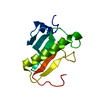
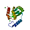
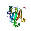
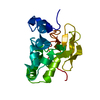
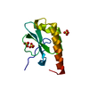
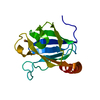
| Title | Crystal structure of the C-terminal MA3 domain of Pdcd4 (mouse); form 3 | ||||||
 Components Components | Programmed Cell Death 4, Pdcd4 | ||||||
 Keywords Keywords | ANTITUMOR PROTEIN / alpha-helical | ||||||
| Function / homology |  Function and homology information Function and homology informationepithelial to mesenchymal transition involved in cardiac fibroblast development / negative regulation of myofibroblast differentiation / negative regulation of vascular associated smooth muscle cell differentiation / positive regulation of smooth muscle cell apoptotic process / negative regulation of JUN kinase activity / regulation of protein metabolic process / response to alkaloid / positive regulation of endothelial cell apoptotic process / positive regulation of vascular associated smooth muscle cell apoptotic process / negative regulation of vascular associated smooth muscle cell proliferation ...epithelial to mesenchymal transition involved in cardiac fibroblast development / negative regulation of myofibroblast differentiation / negative regulation of vascular associated smooth muscle cell differentiation / positive regulation of smooth muscle cell apoptotic process / negative regulation of JUN kinase activity / regulation of protein metabolic process / response to alkaloid / positive regulation of endothelial cell apoptotic process / positive regulation of vascular associated smooth muscle cell apoptotic process / negative regulation of vascular associated smooth muscle cell proliferation / negative regulation of cytokine production involved in inflammatory response / BMP signaling pathway / response to hormone / positive regulation of inflammatory response / positive regulation of non-canonical NF-kappaB signal transduction / cellular response to lipopolysaccharide / negative regulation of DNA-templated transcription / apoptotic process / negative regulation of apoptotic process / RNA binding / nucleus / cytosol / cytoplasmSimilarity search - Function | ||||||
| Biological species |  Mus musculus (house mouse) Mus musculus (house mouse) | ||||||
| Method | X-RAY DIFFRACTION / SYNCHROTRON / MOLECULAR REPLACEMENT / Resolution: 1.76 Å | ||||||
 Authors Authors | Wlodawer, A. / LaRonde-LeBlanc, N.A. | ||||||
 Citation Citation | Journal: Mol.Cell.Biol. / Year: 2007 Title: Structural basis for inhibition of translation by the tumor suppressor pdcd4. Authors: Laronde-Leblanc, N. / Santhanam, A.N. / Baker, A.R. / Wlodawer, A. / Colburn, N.H. | ||||||
| History |
|
- Structure visualization
| Structure viewer | Molecule: MolmilJmol/JSmol |
|---|
- Downloads & links
-Download
| PDBx/mmCIF format | 2ios.cif.gz | 41.6 KB | Display | PDBx/mmCIF format |
|---|---|---|---|---|
| PDB format | pdb2ios.ent.gz | 27.8 KB | Display | PDB format |
| PDBx/mmJSON format | 2ios.json.gz | Tree view | PDBx/mmJSON format | |
| Others |  Other downloads Other downloads |
-Validation report
| Arichive directory | https://data.pdbj.org/pub/pdb/validation_reports/io/2iosftp://data.pdbj.org/pub/pdb/validation_reports/io/2ios | HTTPS FTP |
|---|
-Related structure data
| Related structure data |  2iolSC 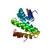 2ionC 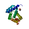 2nszC S: Starting model for refinement C: citing same article ( |
|---|---|
| Similar structure data |


-Links
 PDBj
PDBj
- Assembly
| Deposited unit | 
| |||||||||
|---|---|---|---|---|---|---|---|---|---|---|
| 1 |
| |||||||||
| Unit cell |
| |||||||||
| Components on special symmetry positions |
|
-Components
| #1: Protein | / Pdcd4 Mass: 17350.844 Da / Num. of mol.: 1 / Fragment: C-terminal MA3 domain, residues 323-448 Source method: isolated from a genetically manipulated source Source: (gene. exp.) Mus musculus (house mouse) / Gene: Pdcd4, MA-3 / Plasmid: pDEST14 / Production host:  Escherichia coli (E. coli) / Strain (production host): Rosetta(DE3)pLysS / References: UniProt: Q61823 Escherichia coli (E. coli) / Strain (production host): Rosetta(DE3)pLysS / References: UniProt: Q61823 |
|---|---|
| #2: Water | ChemComp-HOH / Water Mass: 18.015 Da / Num. of mol.: 116 / Source method: isolated from a natural source / Formula: H2O Mass: 18.015 Da / Num. of mol.: 116 / Source method: isolated from a natural source / Formula: H2O |
-Experimental details
-Experiment
| Experiment | Method: X-RAY DIFFRACTION / Number of used crystals: 1 |
|---|
- Sample preparation
| Crystal | Density Matthews: 2.61 Å3/Da / Density % sol: 52.92 % |
|---|---|
| Crystal grow | Temperature: 293 K / Method: vapor diffusion, hanging drop / pH: 4.2 Details: 10% (w/v) PEG-3000, 100 mM phosphate-citrate, 200 mM NaCl, pH 4.2, VAPOR DIFFUSION, HANGING DROP, temperature 293K |
-Data collection
| Diffraction | Mean temperature: 100 K |
|---|---|
| Diffraction source | Source: SYNCHROTRON / Site: APS  / Beamline: 22-ID / Wavelength: 0.97931 Å / Beamline: 22-ID / Wavelength: 0.97931 Å |
| Detector | Type: MARMOSAIC 300 mm CCD / Detector: CCD |
| Radiation | Protocol: SINGLE WAVELENGTH / Monochromatic (M) / Laue (L): M / Scattering type: x-ray |
| Radiation wavelength | Wavelength: 0.97931 Å / Relative weight: 1 |
| Reflection | Resolution: 1.76→30 Å / Num. all: 15015 / Num. obs: 14854 / % possible obs: 99.5 % / Observed criterion σ(F): 1 / Observed criterion σ(I): 1 / Redundancy: 3.7 % / Rsym value: 0.033 / Net I/σ(I): 32.2 |
| Reflection shell | Resolution: 1.76→1.806 Å / Redundancy: 3.3 % / Mean I/σ(I) obs: 4.54 / Num. unique all: 1034 / Rsym value: 0.248 / % possible all: 95.6 |
- Processing
| Software |
| ||||||||||||||||||||||||||||||||||||||||||||||||||||||||||||||||||||||||||||||||||||||||||
|---|---|---|---|---|---|---|---|---|---|---|---|---|---|---|---|---|---|---|---|---|---|---|---|---|---|---|---|---|---|---|---|---|---|---|---|---|---|---|---|---|---|---|---|---|---|---|---|---|---|---|---|---|---|---|---|---|---|---|---|---|---|---|---|---|---|---|---|---|---|---|---|---|---|---|---|---|---|---|---|---|---|---|---|---|---|---|---|---|---|---|---|
| Refinement | Method to determine structure: MOLECULAR REPLACEMENT Starting model: PDB ENTRY 2IOL, chain A. Resolution: 1.76→30 Å / Cor.coef. Fo:Fc: 0.963 / Cor.coef. Fo:Fc free: 0.93 / SU B: 2.584 / SU ML: 0.085 / Isotropic thermal model: Isotropic / Cross valid method: THROUGHOUT / σ(F): 0 / σ(I): 0 / ESU R: 0.119 / ESU R Free: 0.126 / Stereochemistry target values: MAXIMUM LIKELIHOOD
| ||||||||||||||||||||||||||||||||||||||||||||||||||||||||||||||||||||||||||||||||||||||||||
| Solvent computation | Ion probe radii: 0.8 Å / Shrinkage radii: 0.8 Å / VDW probe radii: 1.2 Å / Solvent model: MASK | ||||||||||||||||||||||||||||||||||||||||||||||||||||||||||||||||||||||||||||||||||||||||||
| Displacement parameters | Biso mean: 32.106 Å2
| ||||||||||||||||||||||||||||||||||||||||||||||||||||||||||||||||||||||||||||||||||||||||||
| Refine analyze |
| ||||||||||||||||||||||||||||||||||||||||||||||||||||||||||||||||||||||||||||||||||||||||||
| Refinement step | Cycle: LAST / Resolution: 1.76→30 Å
| ||||||||||||||||||||||||||||||||||||||||||||||||||||||||||||||||||||||||||||||||||||||||||
| Refine LS restraints |
| ||||||||||||||||||||||||||||||||||||||||||||||||||||||||||||||||||||||||||||||||||||||||||
| LS refinement shell | Resolution: 1.76→1.806 Å / Total num. of bins used: 20
|
