 Movie
Movie Controller


+ Open data

- Basic information
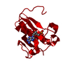
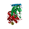
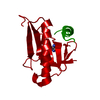
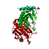
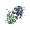
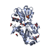
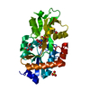
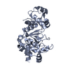
| Entry | Database: PDB / ID: 4fit | ||||||
|---|---|---|---|---|---|---|---|
| Title | FHIT-APO | ||||||
 Components Components | FRAGILE HISTIDINE TRIAD PROTEIN FHIT FHIT | ||||||
 Keywords Keywords | HYDROLASE / FRAGILE HISTIDINE TRIAD PROTEIN / FHIT / PUTATIVE TUMOR SUPPRESSOR / HIT PROTEIN FAMILY / HISTIDINE TRIAD PROTEIN FAMILY / NUCLEOTIDYL HYDROLASE / NUCLEOTIDYL TRANSFERASE | ||||||
| Function / homology |  Function and homology informationadenylylsulfate-ammonia adenylyltransferase / adenylylsulfatase / diadenosine triphosphate catabolic process / adenylylsulfate-ammonia adenylyltransferase activity / adenylylsulfatase activity / bis(5'-adenosyl)-triphosphatase / bis(5'-adenosyl)-triphosphatase activity / purine nucleotide metabolic process / Hydrolases; Acting on phosphorus-nitrogen bonds / adenosine 5'-monophosphoramidase activity ...adenylylsulfate-ammonia adenylyltransferase / adenylylsulfatase / diadenosine triphosphate catabolic process / adenylylsulfate-ammonia adenylyltransferase activity / adenylylsulfatase activity / bis(5'-adenosyl)-triphosphatase / bis(5'-adenosyl)-triphosphatase activity / purine nucleotide metabolic process / Hydrolases; Acting on phosphorus-nitrogen bonds / adenosine 5'-monophosphoramidase activity / intrinsic apoptotic signaling pathway by p53 class mediator / negative regulation of proteasomal ubiquitin-dependent protein catabolic process / fibrillar center / nucleotide binding / ubiquitin protein ligase binding / mitochondrion / identical protein binding / nucleus / plasma membrane / cytosol / cytoplasm Function and homology informationadenylylsulfate-ammonia adenylyltransferase / adenylylsulfatase / diadenosine triphosphate catabolic process / adenylylsulfate-ammonia adenylyltransferase activity / adenylylsulfatase activity / bis(5'-adenosyl)-triphosphatase / bis(5'-adenosyl)-triphosphatase activity / purine nucleotide metabolic process / Hydrolases; Acting on phosphorus-nitrogen bonds / adenosine 5'-monophosphoramidase activity ...adenylylsulfate-ammonia adenylyltransferase / adenylylsulfatase / diadenosine triphosphate catabolic process / adenylylsulfate-ammonia adenylyltransferase activity / adenylylsulfatase activity / bis(5'-adenosyl)-triphosphatase / bis(5'-adenosyl)-triphosphatase activity / purine nucleotide metabolic process / Hydrolases; Acting on phosphorus-nitrogen bonds / adenosine 5'-monophosphoramidase activity / intrinsic apoptotic signaling pathway by p53 class mediator / negative regulation of proteasomal ubiquitin-dependent protein catabolic process / fibrillar center / nucleotide binding / ubiquitin protein ligase binding / mitochondrion / identical protein binding / nucleus / plasma membrane / cytosol / cytoplasmSimilarity search - Function | ||||||
| Biological species |  Homo sapiens (human) Homo sapiens (human) | ||||||
| Method | X-RAY DIFFRACTION / Resolution: 2.5 Å | ||||||
 Authors Authors | Lima, C.D. / Klein, M.G. / Hendrickson, W.A. | ||||||
 Citation Citation | Journal: Science / Year: 1997 Title: Structure-based analysis of catalysis and substrate definition in the HIT protein family. Authors: Lima, C.D. / Klein, M.G. / Hendrickson, W.A. #1: Journal: Structure / Year: 1997Title: MAD Analysis of Fhit, a Putative Human Tumor Suppressor from the Hit Protein Family Authors: Lima, C.D. / D'Amico, K.L. / Naday, I. / Rosenbaum, G. / Westbrook, E.M. / Hendrickson, W.A. | ||||||
| History |
|
- Structure visualization
| Structure viewer | Molecule: MolmilJmol/JSmol |
|---|
- Downloads & links
-Download
| PDBx/mmCIF format | 4fit.cif.gz | 37.5 KB | Display | PDBx/mmCIF format |
|---|---|---|---|---|
| PDB format | pdb4fit.ent.gz | 25.9 KB | Display | PDB format |
| PDBx/mmJSON format | 4fit.json.gz | Tree view | PDBx/mmJSON format | |
| Others |  Other downloads Other downloads |
-Validation report
| Arichive directory | https://data.pdbj.org/pub/pdb/validation_reports/fi/4fitftp://data.pdbj.org/pub/pdb/validation_reports/fi/4fit | HTTPS FTP |
|---|
-Related structure data


-Links
 PDBj
PDBj


- Assembly
| Deposited unit | 
| ||||||||
|---|---|---|---|---|---|---|---|---|---|
| 1 | 
| ||||||||
| Unit cell |
|
-Components
| #1: Protein | FHIT / FHIT Mass: 16886.203 Da / Num. of mol.: 1 Source method: isolated from a genetically manipulated source Source: (gene. exp.) Homo sapiens (human)Description: EXPRESSED AS FUSION PROTEIN WITH GLUTATHIONE-S-TRANSFERASE IN ESCHERICHIA COLI Gene: FHIT / Plasmid: PGEX-2T / Gene (production host): FHIT / Production host:  Escherichia coli (E. coli) / Strain (production host): DH5-ALPHA Escherichia coli (E. coli) / Strain (production host): DH5-ALPHAReferences: UniProt: P49789, bis(5'-adenosyl)-triphosphatase |
|---|---|
| #2: Water | ChemComp-HOH / Water Mass: 18.015 Da / Num. of mol.: 67 / Source method: isolated from a natural source / Formula: H2O Mass: 18.015 Da / Num. of mol.: 67 / Source method: isolated from a natural source / Formula: H2O |
-Experimental details
-Experiment
| Experiment | Method: X-RAY DIFFRACTION / Number of used crystals: 1 |
|---|
- Sample preparation
| Crystal | Density Matthews: 2.93 Å3/Da / Density % sol: 58.01 % | ||||||||||||||||||||
|---|---|---|---|---|---|---|---|---|---|---|---|---|---|---|---|---|---|---|---|---|---|
| Crystal grow | pH: 6.5 / Details: GROWN FROM AMMONIUM SULFATE, PH 6.5 | ||||||||||||||||||||
| Crystal grow | *PLUS Method: unknown / Details: Lima, C.D., (1997) Structure (London), 5, 763. | ||||||||||||||||||||
| Components of the solutions | *PLUS
|
-Data collection
| Diffraction | Mean temperature: 110 K |
|---|---|
| Diffraction source | Wavelength: 1.5418 |
| Detector | Type: RIGAKU / Detector: IMAGE PLATE / Date: Dec 16, 1996 |
| Radiation | Monochromatic (M) / Laue (L): M / Scattering type: x-ray |
| Radiation wavelength | Wavelength: 1.5418 Å / Relative weight: 1 |
| Reflection | Resolution: 2.3→20 Å / Num. obs: 16062 / % possible obs: 94.6 % / Observed criterion σ(I): 2 / Redundancy: 8 % / Rmerge(I) obs: 0.076 |
| Reflection | *PLUS Num. measured all: 100846 |
- Processing
| Software |
| ||||||||||||||||||||||||||||||||||||||||||||||||||||||||||||
|---|---|---|---|---|---|---|---|---|---|---|---|---|---|---|---|---|---|---|---|---|---|---|---|---|---|---|---|---|---|---|---|---|---|---|---|---|---|---|---|---|---|---|---|---|---|---|---|---|---|---|---|---|---|---|---|---|---|---|---|---|---|
| Refinement | Resolution: 2.5→8 Å / Data cutoff high absF: 100000 / Data cutoff low absF: 0.1 / σ(F): 2
| ||||||||||||||||||||||||||||||||||||||||||||||||||||||||||||
| Displacement parameters | Biso mean: 29.3 Å2 | ||||||||||||||||||||||||||||||||||||||||||||||||||||||||||||
| Refinement step | Cycle: LAST / Resolution: 2.5→8 Å
| ||||||||||||||||||||||||||||||||||||||||||||||||||||||||||||
| Refine LS restraints |
| ||||||||||||||||||||||||||||||||||||||||||||||||||||||||||||
| Software | *PLUS Name: X-PLOR / Version: 3.1 / Classification: refinement | ||||||||||||||||||||||||||||||||||||||||||||||||||||||||||||
| Refine LS restraints | *PLUS
|
