 Movie
Movie Controller

[English] 日本語
 Yorodumi
- PDB-2inw: Crystal structure of Q83JN9 from Shigella flexneri at high resolu... -
+ Open data

- Basic information
| Entry | Database: PDB / ID: 2inw | ||||||
|---|---|---|---|---|---|---|---|
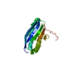
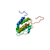
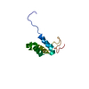
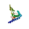
| Title | Crystal structure of Q83JN9 from Shigella flexneri at high resolution. Northeast Structural Genomics Consortium target SfR137. | ||||||
 Components Components | Putative structural protein | ||||||
 Keywords Keywords | STRUCTURAL GENOMICS / UNKNOWN FUNCTION / Q83JN9 X-Ray NESG SfR137 / PSI-2 / Protein Structure Initiative / Northeast Structural Genomics Consortium | ||||||
| Function / homology |  Function and homology information Function and homology information | ||||||
| Biological species |  Shigella flexneri (bacteria) Shigella flexneri (bacteria) | ||||||
| Method |  X-RAY DIFFRACTION / SYNCHROTRON / SAD / Resolution: 1.5 Å X-RAY DIFFRACTION / SYNCHROTRON / SAD / Resolution: 1.5 Å | ||||||
 Authors Authors | Kuzin, A.P. / Su, M. / Jayaraman, S. / Vorobiev, S.M. / Wang, D. / Jiang, M. / Cunningham, K. / Ma, L.-C. / Xiao, R. / Liu, J. ...Kuzin, A.P. / Su, M. / Jayaraman, S. / Vorobiev, S.M. / Wang, D. / Jiang, M. / Cunningham, K. / Ma, L.-C. / Xiao, R. / Liu, J. / Baran, M. / Swapna, G.V.T. / Acton, T.B. / Rost, B. / Montelione, G.T. / Tong, L. / Hunt, J.F. / Northeast Structural Genomics Consortium (NESG) | ||||||
 Citation Citation | Journal: Structure / Year: 2010 Title: Crystal Structures of Phd-Doc, HigA, and YeeU Establish Multiple Evolutionary Links between Microbial Growth-Regulating Toxin-Antitoxin Systems. Authors: Arbing, M.A. / Handelman, S.K. / Kuzin, A.P. / Verdon, G. / Wang, C. / Su, M. / Rothenbacher, F.P. / Abashidze, M. / Liu, M. / Hurley, J.M. / Xiao, R. / Acton, T. / Inouye, M. / Montelione, ...Authors: Arbing, M.A. / Handelman, S.K. / Kuzin, A.P. / Verdon, G. / Wang, C. / Su, M. / Rothenbacher, F.P. / Abashidze, M. / Liu, M. / Hurley, J.M. / Xiao, R. / Acton, T. / Inouye, M. / Montelione, G.T. / Woychik, N.A. / Hunt, J.F. | ||||||
| History |
|
- Structure visualization
| Structure viewer | Molecule: MolmilJmol/JSmol |
|---|
- Downloads & links
-Download
| PDBx/mmCIF format | 2inw.cif.gz | 60.1 KB | Display | PDBx/mmCIF format |
|---|---|---|---|---|
| PDB format | pdb2inw.ent.gz | 47.5 KB | Display | PDB format |
| PDBx/mmJSON format | 2inw.json.gz | Tree view | PDBx/mmJSON format | |
| Others |  Other downloads Other downloads |
-Validation report
| Summary document | 2inw_validation.pdf.gz | 439 KB | Display | wwPDB validaton report |
|---|---|---|---|---|
| Full document | 2inw_full_validation.pdf.gz | 440.9 KB | Display | |
| Data in XML | 2inw_validation.xml.gz | 13.6 KB | Display | |
| Data in CIF | 2inw_validation.cif.gz | 19.6 KB | Display | |
| Arichive directory | https://data.pdbj.org/pub/pdb/validation_reports/in/2inwftp://data.pdbj.org/pub/pdb/validation_reports/in/2inw | HTTPS FTP |
-Related structure data
| Related structure data |  2h28C  2ictC  3kh2C C: citing same article ( |
|---|---|
| Similar structure data | |
| Other databases |


-Links
 PDBj
PDBj- Assembly
| Deposited unit | 
| ||||||||
|---|---|---|---|---|---|---|---|---|---|
| 1 | 
| ||||||||
| 2 | 
| ||||||||
| Unit cell |
|
-Components
| #1: Protein | Mass: 14956.386 Da / Num. of mol.: 2 Source method: isolated from a genetically manipulated source Source: (gene. exp.) Shigella flexneri (bacteria) / Gene: yeeU, SF2998, S_3203 / Plasmid: pET21 / Production host: #2: Chemical | ChemComp-PO4 / |   Mass: 94.971 Da / Num. of mol.: 1 / Source method: obtained synthetically / Formula: PO4 Mass: 94.971 Da / Num. of mol.: 1 / Source method: obtained synthetically / Formula: PO4#3: Water | ChemComp-HOH / |  Mass: 18.015 Da / Num. of mol.: 252 / Source method: isolated from a natural source / Formula: H2O Mass: 18.015 Da / Num. of mol.: 252 / Source method: isolated from a natural source / Formula: H2O |
|---|
-Experimental details
-Experiment
| Experiment | Method: X-RAY DIFFRACTION / Number of used crystals: 1 |
|---|
- Sample preparation
| Crystal | Density Matthews: 2.15 Å3/Da / Density % sol: 42.9 % |
|---|---|
| Crystal grow | Temperature: 293 K / Method: vapor diffusion, hanging drop / pH: 6 Details: 0.1M Kh2PO4, 0.1M MES, 20% PEG20K, pH 6.0, VAPOR DIFFUSION, HANGING DROP, temperature 293K |
-Data collection
| Diffraction source | Source: SYNCHROTRON / Site: NSLS  / Beamline: X4A / Wavelength: 0.979 Å / Beamline: X4A / Wavelength: 0.979 Å |
|---|---|
| Detector | Type: ADSC QUANTUM 4 / Detector: CCD / Date: Sep 26, 2006 / Details: mirrors |
| Radiation | Protocol: SINGLE WAVELENGTH / Monochromatic (M) / Laue (L): M / Scattering type: x-ray |
| Radiation wavelength | Wavelength: 0.979 Å / Relative weight: 1 |
| Reflection | Resolution: 1.5→30 Å / Num. all: 77681 / Num. obs: 41154 / % possible obs: 97.1 % / Observed criterion σ(I): -3 / Biso Wilson estimate: 17.1 Å2 / Rmerge(I) obs: 0.073 / Net I/σ(I): 24.9 |
| Reflection shell | Resolution: 1.5→1.55 Å / Rmerge(I) obs: 0.65 / Mean I/σ(I) obs: 2 / % possible all: 79.8 |
- Processing
| Software |
| ||||||||||||||||||||
|---|---|---|---|---|---|---|---|---|---|---|---|---|---|---|---|---|---|---|---|---|---|
| Refinement | Method to determine structure: SAD / Resolution: 1.5→19.42 Å / Rfactor Rfree error: 0.004 / Data cutoff high absF: 144000.44 / Data cutoff low absF: 0 / Isotropic thermal model: RESTRAINED / Cross valid method: THROUGHOUT / σ(F): 1 / Stereochemistry target values: Engh & Huber
| ||||||||||||||||||||
| Solvent computation | Solvent model: FLAT MODEL / Bsol: 38.2607 Å2 / ksol: 0.325375 e/Å3 | ||||||||||||||||||||
| Displacement parameters | Biso mean: 20.2 Å2
| ||||||||||||||||||||
| Refine analyze |
| ||||||||||||||||||||
| Refinement step | Cycle: LAST / Resolution: 1.5→19.42 Å
| ||||||||||||||||||||
| Refine LS restraints |
| ||||||||||||||||||||
| LS refinement shell | Resolution: 1.5→1.59 Å / Rfactor Rfree error: 0.017 / Total num. of bins used: 6
| ||||||||||||||||||||
| Xplor file |
|
