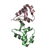
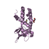
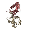
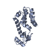
structures with acceptable covalent geometry,structures with the least restraint violations,structures with the lowest energy
Representative
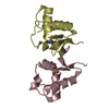
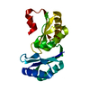
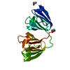
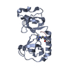
Model #1
lowest energy
-
Components
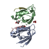
#1: Protein
Thiol:disulfideinterchangeproteindsbA
Mass: 20495.365 Da / Num. of mol.: 1 Source method: isolated from a genetically manipulated source Source: (gene. exp.) Vibrio cholerae (bacteria) / Gene: dsbA, tpcG / Plasmid: pET / Species (production host): Escherichia coli / Production host: Escherichia coli BL21(DE3) (bacteria) / Strain (production host): BL21-DE3 References: UniProt: P32557, protein-disulfide reductase (glutathione)
-
Experimental details
-
Experiment
Experiment
Method: SOLUTION NMR
NMR experiment
Conditions-ID
Experiment-ID
Solution-ID
Type
1
1
1
3D 13C-separated NOESY
1
2
1
3D 15N-separated NOESY
NMR details
Text: Structure determined using triple resonance methods. Stereospecific assignments of valine and leucine methyls were achieved using a 10% 13C-labeled sample of DsbA
-
Sample preparation
Details
Solution-ID
Contents
Solvent system
1
400 uM DsbA U-15N,13C, 10 mM HEPES, 50 mM sodium chloride, ph 6.8, 90% H2O, 10% D2O
90% H2O/10% D2O
2
400 uM DsbA 10%-13C, 10 mM HEPES, 50 mM sodium chloride, ph 6.8, 90% H2O, 10% D2O
90% H2O/10% D2O
Sample conditions
Ionic strength: 50 mM NaCl / pH: 6.8 / Pressure: 1 atm / Temperature: 320 K
-
NMR measurement
Radiation
Protocol: SINGLE WAVELENGTH / Monochromatic (M) / Laue (L): M
Radiation wavelength
Relative weight: 1
NMR spectrometer
Type
Manufacturer
Model
Field strength (MHz)
Spectrometer-ID
Bruker DRX
Bruker
DRX
500
1
Varian AVANCE
Varian
AVANCE
600
2
-
Processing
NMR software
Name
Version
Classification
XwinNMR
collection
VNMR
collection
NMRPipe
processing
Sparky
dataanalysis
CYANA
2.1
structuresolution
X-PLOR
refinement
Refinement
Method: torsion angle dynamics / Software ordinal: 1 Details: 898 long range NOE restraints, 850 medium range NOE restraints, 95 ambiguous NOE restraints, 95 hydrogen bond distance restraints, 238 dihedral angle restraints
NMR representative
Selection criteria: lowest energy
NMR ensemble
Conformer selection criteria: structures with acceptable covalent geometry,structures with the least restraint violations,structures with the lowest energy Conformers calculated total number: 100 / Conformers submitted total number: 22
+
About Yorodumi
-
News
-
Feb 9, 2022. New format data for meta-information of EMDB entries
New format data for meta-information of EMDB entries
Version 3 of the EMDB header file is now the official format.
The previous official version 1.9 will be removed from the archive.
In the structure databanks used in Yorodumi, some data are registered as the other names, "COVID-19 virus" and "2019-nCoV". Here are the details of the virus and the list of structure data.
Jan 31, 2019. EMDB accession codes are about to change! (news from PDBe EMDB page)
EMDB accession codes are about to change! (news from PDBe EMDB page)
The allocation of 4 digits for EMDB accession codes will soon come to an end. Whilst these codes will remain in use, new EMDB accession codes will include an additional digit and will expand incrementally as the available range of codes is exhausted. The current 4-digit format prefixed with “EMD-” (i.e. EMD-XXXX) will advance to a 5-digit format (i.e. EMD-XXXXX), and so on. It is currently estimated that the 4-digit codes will be depleted around Spring 2019, at which point the 5-digit format will come into force.
The EM Navigator/Yorodumi systems omit the EMD- prefix.
Related info.:Q: What is EMD? / ID/Accession-code notation in Yorodumi/EM Navigator
Yorodumi is a browser for structure data from EMDB, PDB, SASBDB, etc.
This page is also the successor to EM Navigator detail page, and also detail information page/front-end page for Omokage search.
The word "yorodu" (or yorozu) is an old Japanese word meaning "ten thousand". "mi" (miru) is to see.
Related info.:EMDB / PDB / SASBDB / Comparison of 3 databanks / Yorodumi Search / Aug 31, 2016. New EM Navigator & Yorodumi / Yorodumi Papers / Jmol/JSmol / Function and homology information / Changes in new EM Navigator and Yorodumi
 Movie
Movie Controller

 Yorodumi
 Open data
 Basic information
 Components
 Keywords
 OXIDOREDUCTASE /
  Function and homology information

 Authors
 Citation
 Structure visualization
 Downloads & links
 Other downloads
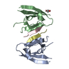
 PDBj
PDBj Assembly
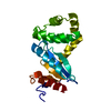
 Sample preparation
 Processing