 Movie
Movie Controller

[English] 日本語
 Yorodumi
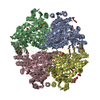
- PDB-2ihv: Carboxyethylarginine synthase from Streptomyces clavuligerus: 5-g... -
+ Open data

- Basic information
| Entry | Database: PDB / ID: 2ihv | ||||||
|---|---|---|---|---|---|---|---|
| Title | Carboxyethylarginine synthase from Streptomyces clavuligerus: 5-guanidinovaleric acid complex | ||||||
 Components Components | Carboxyethylarginine synthase | ||||||
 Keywords Keywords |  TRANSFERASE / Thiamin diphosphate complex TRANSFERASE / Thiamin diphosphate complex | ||||||
| Function / homology |  Function and homology informationN2-(2-carboxyethyl)arginine synthase / N2-(2-carboxyethyl)arginine synthase activity / thiamine pyrophosphate binding / magnesium ion binding Function and homology informationN2-(2-carboxyethyl)arginine synthase / N2-(2-carboxyethyl)arginine synthase activity / thiamine pyrophosphate binding / magnesium ion bindingSimilarity search - Function | ||||||
| Biological species |  Streptomyces clavuligerus (bacteria) Streptomyces clavuligerus (bacteria) | ||||||
| Method | X-RAY DIFFRACTION / SYNCHROTRON / MOLECULAR REPLACEMENT / Resolution: 2.3 Å | ||||||
 Authors Authors | Caines, M.E. / Schofield, C.J. | ||||||
 Citation Citation | Journal: Biochem.Biophys.Res.Commun. / Year: 2009 Title: Structural and mechanistic studies on N(2)-(2-carboxyethyl)arginine synthase. Authors: Caines, M.E. / Sorensen, J.L. / Schofield, C.J. | ||||||
| History |
|
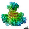
- Structure visualization
| Structure viewer | Molecule: MolmilJmol/JSmol |
|---|
- Downloads & links
-Download
| PDBx/mmCIF format | 2ihv.cif.gz | 444.8 KB | Display | PDBx/mmCIF format |
|---|---|---|---|---|
| PDB format | pdb2ihv.ent.gz | 359.2 KB | Display | PDB format |
| PDBx/mmJSON format | 2ihv.json.gz | Tree view | PDBx/mmJSON format | |
| Others |  Other downloads Other downloads |
-Validation report
| Arichive directory | https://data.pdbj.org/pub/pdb/validation_reports/ih/2ihvftp://data.pdbj.org/pub/pdb/validation_reports/ih/2ihv | HTTPS FTP |
|---|
-Related structure data
| Related structure data |  2ihtC  2ihuC  1upaS S: Starting model for refinement C: citing same article ( |
|---|---|
| Similar structure data |


-Links
 PDBj
PDBj


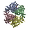
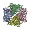
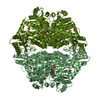
- Assembly
| Deposited unit | 
| |||||||||||||||||||||||||||||||||||||||||||||||||||||||||||||||||||||||||||||||||||||||||||||||||||||||||||||||||||||
|---|---|---|---|---|---|---|---|---|---|---|---|---|---|---|---|---|---|---|---|---|---|---|---|---|---|---|---|---|---|---|---|---|---|---|---|---|---|---|---|---|---|---|---|---|---|---|---|---|---|---|---|---|---|---|---|---|---|---|---|---|---|---|---|---|---|---|---|---|---|---|---|---|---|---|---|---|---|---|---|---|---|---|---|---|---|---|---|---|---|---|---|---|---|---|---|---|---|---|---|---|---|---|---|---|---|---|---|---|---|---|---|---|---|---|---|---|---|---|
| 1 |
| |||||||||||||||||||||||||||||||||||||||||||||||||||||||||||||||||||||||||||||||||||||||||||||||||||||||||||||||||||||
| Unit cell |
| |||||||||||||||||||||||||||||||||||||||||||||||||||||||||||||||||||||||||||||||||||||||||||||||||||||||||||||||||||||
| Noncrystallographic symmetry (NCS) | NCS domain:
NCS domain segments: Ens-ID: 1 / Refine code: 4
|
-Components
-Protein , 1 types, 4 molecules ABCD
| #1: Protein | Mass: 60964.828 Da / Num. of mol.: 4 Source method: isolated from a genetically manipulated source Source: (gene. exp.) Streptomyces clavuligerus (bacteria) / Gene: ceas2 / Plasmid: pET24a(+) / Species (production host): Escherichia coli / Production host: Escherichia coli BL21(DE3) (bacteria) / Strain (production host): BL21(DE3)References: UniProt: Q9LCV9, N2-(2-carboxyethyl)arginine synthase |
|---|
-Non-polymers , 5 types, 1008 molecules 


| #2: Chemical | ChemComp-MG /  Mass: 24.305 Da / Num. of mol.: 4 / Source method: obtained synthetically / Formula: Mg Mass: 24.305 Da / Num. of mol.: 4 / Source method: obtained synthetically / Formula: Mg#3: Chemical | ChemComp-K /  Mass: 39.098 Da / Num. of mol.: 6 / Source method: obtained synthetically / Formula: K Mass: 39.098 Da / Num. of mol.: 6 / Source method: obtained synthetically / Formula: K#4: Chemical | ChemComp-TPP / Thiamine pyrophosphate Mass: 425.314 Da / Num. of mol.: 4 / Source method: obtained synthetically / Formula: C12H19N4O7P2S Mass: 425.314 Da / Num. of mol.: 4 / Source method: obtained synthetically / Formula: C12H19N4O7P2S#5: Chemical | ChemComp-GVA /  Mass: 159.186 Da / Num. of mol.: 4 / Source method: obtained synthetically / Formula: C6H13N3O2 Mass: 159.186 Da / Num. of mol.: 4 / Source method: obtained synthetically / Formula: C6H13N3O2#6: Water | ChemComp-HOH / | WaterMass: 18.015 Da / Num. of mol.: 990 / Source method: isolated from a natural source / Formula: H2O |
|---|
-Experimental details
-Experiment
| Experiment | Method: X-RAY DIFFRACTION / Number of used crystals: 1 |
|---|
- Sample preparation
| Crystal | Density Matthews: 3.14 Å3/Da / Density % sol: 60.79 % |
|---|---|
| Crystal grow | Temperature: 290 K / Method: vapor diffusion, hanging drop / pH: 7.4 Details: 1.6 M (NH4)2SO4, 0.1 M HEPES pH 7.4, 10 mg/mL protein with 3-fold excess of ThDP, VAPOR DIFFUSION, HANGING DROP, temperature 290K |
-Data collection
| Diffraction | Mean temperature: 100 K |
|---|---|
| Diffraction source | Source: SYNCHROTRON / Site: SRS  / Beamline: PX10.1 / Wavelength: 1.488 Å / Beamline: PX10.1 / Wavelength: 1.488 Å |
| Detector | Type: MAR CCD 225 mm / Detector: CCD / Date: Nov 10, 2004 |
| Radiation | Monochromator: Si(III) / Protocol: SINGLE WAVELENGTH / Monochromatic (M) / Laue (L): M / Scattering type: x-ray |
| Radiation wavelength | Wavelength: 1.488 Å / Relative weight: 1 |
| Reflection | Resolution: 2.3→50 Å / Num. all: 136572 / Num. obs: 131792 / % possible obs: 96.5 % / Observed criterion σ(F): 0 / Observed criterion σ(I): 0 / Redundancy: 5 % / Biso Wilson estimate: 30.122 Å2 / Rmerge(I) obs: 0.115 / Χ2: 0.965 / Net I/σ(I): 10.2 |
| Reflection shell | Resolution: 2.3→2.38 Å / Redundancy: 4.4 % / Rmerge(I) obs: 0.347 / Num. unique all: 12059 / Χ2: 0.976 / % possible all: 89.3 |
- Processing
| Software |
| |||||||||||||||||||||||||||||||||||||||||||||||||||||||||||||||||||||||||||||||||||||||||||||||
|---|---|---|---|---|---|---|---|---|---|---|---|---|---|---|---|---|---|---|---|---|---|---|---|---|---|---|---|---|---|---|---|---|---|---|---|---|---|---|---|---|---|---|---|---|---|---|---|---|---|---|---|---|---|---|---|---|---|---|---|---|---|---|---|---|---|---|---|---|---|---|---|---|---|---|---|---|---|---|---|---|---|---|---|---|---|---|---|---|---|---|---|---|---|---|---|---|
| Refinement | Method to determine structure: MOLECULAR REPLACEMENT Starting model: PDB ENTRY 1UPA Resolution: 2.3→49.15 Å / Cor.coef. Fo:Fc: 0.959 / Cor.coef. Fo:Fc free: 0.94 / SU B: 4.618 / SU ML: 0.113 / Cross valid method: THROUGHOUT / σ(F): 0 / σ(I): 0 / ESU R: 0.218 / ESU R Free: 0.171 / Stereochemistry target values: MAXIMUM LIKELIHOOD
| |||||||||||||||||||||||||||||||||||||||||||||||||||||||||||||||||||||||||||||||||||||||||||||||
| Solvent computation | Ion probe radii: 0.8 Å / Shrinkage radii: 0.8 Å / VDW probe radii: 1.4 Å / Solvent model: MASK | |||||||||||||||||||||||||||||||||||||||||||||||||||||||||||||||||||||||||||||||||||||||||||||||
| Displacement parameters | Biso mean: 23.165 Å2
| |||||||||||||||||||||||||||||||||||||||||||||||||||||||||||||||||||||||||||||||||||||||||||||||
| Refinement step | Cycle: LAST / Resolution: 2.3→49.15 Å
| |||||||||||||||||||||||||||||||||||||||||||||||||||||||||||||||||||||||||||||||||||||||||||||||
| Refine LS restraints |
| |||||||||||||||||||||||||||||||||||||||||||||||||||||||||||||||||||||||||||||||||||||||||||||||
| Refine LS restraints NCS | Ens-ID: 1 / Number: 3902 / Refine-ID: X-RAY DIFFRACTION
| |||||||||||||||||||||||||||||||||||||||||||||||||||||||||||||||||||||||||||||||||||||||||||||||
| LS refinement shell | Resolution: 2.3→2.36 Å / Total num. of bins used: 20
|
