 Movie
Movie Controller

[English] 日本語
 Yorodumi
- PDB-2idj: Crystal Structure of Rat Glycine N-Methyltransferase Apoprotein, ... -
+ Open data

- Basic information
| Entry | Database: PDB / ID: 2idj | ||||||
|---|---|---|---|---|---|---|---|
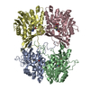
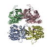
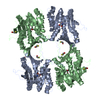
| Title | Crystal Structure of Rat Glycine N-Methyltransferase Apoprotein, Monoclinic Form | ||||||
 Components Components | Glycine N-methyltransferase | ||||||
 Keywords Keywords | TRANSFERASE / Glycine N-methyltransferase / rat / apoprotein. | ||||||
| Function / homology |  Function and homology information Function and homology informationselenol Se-methyltransferase activity / Glyoxylate metabolism and glycine degradation / glycine N-methyltransferase / glycine N-methyltransferase activity / sarcosine metabolic process / methyltransferase complex / methionine metabolic process / S-adenosylhomocysteine metabolic process / glycine metabolic process / S-adenosylmethionine metabolic process ...selenol Se-methyltransferase activity / Glyoxylate metabolism and glycine degradation / glycine N-methyltransferase / glycine N-methyltransferase activity / sarcosine metabolic process / methyltransferase complex / methionine metabolic process / S-adenosylhomocysteine metabolic process / glycine metabolic process / S-adenosylmethionine metabolic process / S-adenosylmethionine-dependent methyltransferase activity / S-adenosyl-L-methionine binding / folic acid binding / glycine binding / regulation of gluconeogenesis / glycogen metabolic process / one-carbon metabolic process / methylation / protein homotetramerization / identical protein binding / cytosolSimilarity search - Function | ||||||
| Biological species |  Rattus norvegicus (Norway rat) Rattus norvegicus (Norway rat) | ||||||
| Method | X-RAY DIFFRACTION / SYNCHROTRON / MOLECULAR REPLACEMENT / Resolution: 2.35 Å | ||||||
 Authors Authors | Luka, Z. / Pakhomova, S. / Loukachevitch, L.V. / Egli, M. / Newcomer, M.E. / Wagner, C. | ||||||
 Citation Citation | Journal: J.Biol.Chem. / Year: 2007 Title: 5-methyltetrahydrofolate is bound in intersubunit areas of rat liver folate-binding protein glycine N-methyltransferase. Authors: Luka, Z. / Pakhomova, S. / Loukachevitch, L.V. / Egli, M. / Newcomer, M.E. / Wagner, C. | ||||||
| History |
|
- Structure visualization
| Structure viewer | Molecule: MolmilJmol/JSmol |
|---|
- Downloads & links
-Download
| PDBx/mmCIF format | 2idj.cif.gz | 228.5 KB | Display | PDBx/mmCIF format |
|---|---|---|---|---|
| PDB format | pdb2idj.ent.gz | 184.1 KB | Display | PDB format |
| PDBx/mmJSON format | 2idj.json.gz | Tree view | PDBx/mmJSON format | |
| Others |  Other downloads Other downloads |
-Validation report
| Arichive directory | https://data.pdbj.org/pub/pdb/validation_reports/id/2idjftp://data.pdbj.org/pub/pdb/validation_reports/id/2idj | HTTPS FTP |
|---|
-Related structure data
| Related structure data | 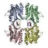 2idkC 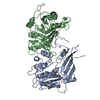 1bhjS S: Starting model for refinement C: citing same article ( |
|---|---|
| Similar structure data |


-Links
 PDBj
PDBj- Assembly
| Deposited unit | 
| ||||||||
|---|---|---|---|---|---|---|---|---|---|
| 1 |
| ||||||||
| Unit cell |
|
-Components
| #1: Protein | / Folate-binding protein Mass: 32460.830 Da / Num. of mol.: 4 Source method: isolated from a genetically manipulated source Source: (gene. exp.) Rattus norvegicus (Norway rat) / Strain: Sprague-Dawley / Gene: Gnmt / Plasmid: pR6 / Species (production host): Escherichia coli / Production host:  Escherichia coli BL21(DE3) (bacteria) / Strain (production host): BL21(DE3) / References: UniProt: P13255, glycine N-methyltransferase Escherichia coli BL21(DE3) (bacteria) / Strain (production host): BL21(DE3) / References: UniProt: P13255, glycine N-methyltransferase#2: Chemical |   Mass: 40.078 Da / Num. of mol.: 2 / Source method: obtained synthetically / Formula: Ca Mass: 40.078 Da / Num. of mol.: 2 / Source method: obtained synthetically / Formula: Ca#3: Water | ChemComp-HOH / | Water Mass: 18.015 Da / Num. of mol.: 115 / Source method: isolated from a natural source / Formula: H2O Mass: 18.015 Da / Num. of mol.: 115 / Source method: isolated from a natural source / Formula: H2O |
|---|
-Experimental details
-Experiment
| Experiment | Method: X-RAY DIFFRACTION / Number of used crystals: 1 |
|---|
- Sample preparation
| Crystal | Density Matthews: 2.5 Å3/Da / Density % sol: 50.85 % |
|---|---|
| Crystal grow | Temperature: 295 K / Method: vapor diffusion, sitting drop / pH: 7.5 Details: 20% PEG 3350, 0.1 M Ca acetate, 0.025 M Tris, pH 7.5, VAPOR DIFFUSION, SITTING DROP, temperature 295K |
-Data collection
| Diffraction | Mean temperature: 100 K |
|---|---|
| Diffraction source | Source: SYNCHROTRON / Site: APS  / Beamline: 22-BM / Wavelength: 1 Å / Beamline: 22-BM / Wavelength: 1 Å |
| Detector | Type: MARMOSAIC 225 mm CCD / Detector: CCD / Date: Oct 28, 2005 / Details: mirrors |
| Radiation | Monochromator: Graphite / Protocol: SINGLE WAVELENGTH / Monochromatic (M) / Laue (L): M / Scattering type: x-ray |
| Radiation wavelength | Wavelength: 1 Å / Relative weight: 1 |
| Reflection | Resolution: 2.35→50 Å / Num. all: 53782 / Num. obs: 53782 / % possible obs: 99.9 % / Observed criterion σ(I): -3 / Redundancy: 3.5 % / Biso Wilson estimate: 30.7 Å2 / Rsym value: 0.66 / Net I/σ(I): 25 |
| Reflection shell | Resolution: 2.35→2.43 Å / Redundancy: 2.9 % / Mean I/σ(I) obs: 2.9 / Num. unique all: 5328 / Rsym value: 0.456 / % possible all: 99.8 |
- Processing
| Software |
| |||||||||||||||||||||||||
|---|---|---|---|---|---|---|---|---|---|---|---|---|---|---|---|---|---|---|---|---|---|---|---|---|---|---|
| Refinement | Method to determine structure: MOLECULAR REPLACEMENT Starting model: PDB entry 1BHJ Resolution: 2.35→39.11 Å / Isotropic thermal model: restrained / Cross valid method: THROUGHOUT / σ(F): 0 / σ(I): 0 / Stereochemistry target values: Engh & Huber
| |||||||||||||||||||||||||
| Displacement parameters | Biso mean: 50.7 Å2
| |||||||||||||||||||||||||
| Refine analyze |
| |||||||||||||||||||||||||
| Refinement step | Cycle: LAST / Resolution: 2.35→39.11 Å
| |||||||||||||||||||||||||
| Refine LS restraints |
| |||||||||||||||||||||||||
| LS refinement shell | Resolution: 2.35→2.5 Å / Rfactor Rfree error: 0.021
|
