 Movie
Movie Controller

[English] 日本語
 Yorodumi
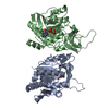
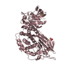
- PDB-1d2g: CRYSTAL STRUCTURE OF R175K MUTANT GLYCINE N-METHYLTRANSFERASE FRO... -
+ Open data

- Basic information
| Entry | Database: PDB / ID: 1d2g | ||||||
|---|---|---|---|---|---|---|---|
| Title | CRYSTAL STRUCTURE OF R175K MUTANT GLYCINE N-METHYLTRANSFERASE FROM RAT LIVER | ||||||
 Components Components | GLYCINE N-METHYLTRANSFERASE | ||||||
 Keywords Keywords | TRANSFERASE / METHYLTRANSFERASE | ||||||
| Function / homology |  Function and homology information Function and homology informationselenol Se-methyltransferase activity / Glyoxylate metabolism and glycine degradation / glycine N-methyltransferase / glycine N-methyltransferase activity / sarcosine metabolic process / methyltransferase complex / methionine metabolic process / S-adenosylhomocysteine metabolic process / glycine metabolic process / S-adenosylmethionine metabolic process ...selenol Se-methyltransferase activity / Glyoxylate metabolism and glycine degradation / glycine N-methyltransferase / glycine N-methyltransferase activity / sarcosine metabolic process / methyltransferase complex / methionine metabolic process / S-adenosylhomocysteine metabolic process / glycine metabolic process / S-adenosylmethionine metabolic process / S-adenosylmethionine-dependent methyltransferase activity / S-adenosyl-L-methionine binding / folic acid binding / glycine binding / regulation of gluconeogenesis / glycogen metabolic process / one-carbon metabolic process / methylation / protein homotetramerization / identical protein binding / cytosolSimilarity search - Function | ||||||
| Biological species |  Rattus norvegicus (Norway rat) Rattus norvegicus (Norway rat) | ||||||
| Method | X-RAY DIFFRACTION / Resolution: 2.5 Å | ||||||
 Authors Authors | Huang, Y. / Komoto, J. / Takusagawa, F. / Konishi, K. / Takata, Y. | ||||||
 Citation Citation | Journal: J.Mol.Biol. / Year: 2000 Title: Mechanisms for auto-inhibition and forced product release in glycine N-methyltransferase: crystal structures of wild-type, mutant R175K and S-adenosylhomocysteine-bound R175K enzymes. Authors: Huang, Y. / Komoto, J. / Konishi, K. / Takata, Y. / Ogawa, H. / Gomi, T. / Fujioka, M. / Takusagawa, F. | ||||||
| History |
|
- Structure visualization
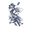
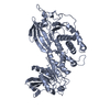
| Structure viewer | Molecule: MolmilJmol/JSmol |
|---|
- Downloads & links
-Download
| PDBx/mmCIF format | 1d2g.cif.gz | 128.7 KB | Display | PDBx/mmCIF format |
|---|---|---|---|---|
| PDB format | pdb1d2g.ent.gz | 102.2 KB | Display | PDB format |
| PDBx/mmJSON format | 1d2g.json.gz | Tree view | PDBx/mmJSON format | |
| Others |  Other downloads Other downloads |
-Validation report
| Arichive directory | https://data.pdbj.org/pub/pdb/validation_reports/d2/1d2gftp://data.pdbj.org/pub/pdb/validation_reports/d2/1d2g | HTTPS FTP |
|---|
-Related structure data


-Links
 PDBj
PDBj- Assembly
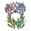
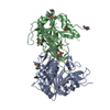
| Deposited unit | 
| ||||||||
|---|---|---|---|---|---|---|---|---|---|
| 1 |
| ||||||||
| 2 | 
| ||||||||
| Unit cell |
| ||||||||
| Details | The biological assembly is a tetramer constructed from chain A and B a symmetry partner generated by the two-fold. |
-Components
| #1: Protein | Mass: 32432.818 Da / Num. of mol.: 2 / Fragment: WHOLE ENZYME / Mutation: R175K Source method: isolated from a genetically manipulated source Source: (gene. exp.) Rattus norvegicus (Norway rat) / Tissue: LIVER / Production host:  Escherichia coli (E. coli) / References: UniProt: P13255, glycine N-methyltransferase Escherichia coli (E. coli) / References: UniProt: P13255, glycine N-methyltransferase#2: Water | ChemComp-HOH / | Water Mass: 18.015 Da / Num. of mol.: 318 / Source method: isolated from a natural source / Formula: H2O Mass: 18.015 Da / Num. of mol.: 318 / Source method: isolated from a natural source / Formula: H2O |
|---|
-Experimental details
-Experiment
| Experiment | Method: X-RAY DIFFRACTION / Number of used crystals: 2 |
|---|
- Sample preparation
| Crystal | Density Matthews: 2.67 Å3/Da / Density % sol: 54.02 % | ||||||||||||||||||||||||||||||||||||
|---|---|---|---|---|---|---|---|---|---|---|---|---|---|---|---|---|---|---|---|---|---|---|---|---|---|---|---|---|---|---|---|---|---|---|---|---|---|
| Crystal grow | Temperature: 277 K / Method: vapor diffusion, hanging drop / pH: 5.6 Details: PEG-4000, pH 5.6, VAPOR DIFFUSION, HANGING DROP, temperature 277K | ||||||||||||||||||||||||||||||||||||
| Crystal grow | *PLUS | ||||||||||||||||||||||||||||||||||||
| Components of the solutions | *PLUS
|
-Data collection
| Diffraction | Mean temperature: 298 K |
|---|---|
| Diffraction source | Source: ROTATING ANODE / Type: RIGAKU RU200 / Wavelength: 1.5418 |
| Detector | Type: MACSCIENCE DIP100 / Detector: IMAGE PLATE / Date: Jan 10, 1997 |
| Radiation | Protocol: SINGLE WAVELENGTH / Monochromatic (M) / Laue (L): M / Scattering type: x-ray |
| Radiation wavelength | Wavelength: 1.5418 Å / Relative weight: 1 |
| Reflection | Resolution: 2.5→10 Å / Num. all: 22390 / Num. obs: 22390 / % possible obs: 91.3 % / Observed criterion σ(F): 0 / Observed criterion σ(I): 0 / Redundancy: 3.3 % / Biso Wilson estimate: 20.8 Å2 / Rmerge(I) obs: 0.071 / Net I/σ(I): 3.35 |
| Reflection shell | Resolution: 2.5→2.6 Å / Redundancy: 2.6 % / Rmerge(I) obs: 0.17 / Num. unique all: 2100 / % possible all: 85 |
| Reflection | *PLUS Lowest resolution: 10 Å / Num. measured all: 71872 |
| Reflection shell | *PLUS Mean I/σ(I) obs: 3.35 |
- Processing
| Software |
| |||||||||||||||||||||||||
|---|---|---|---|---|---|---|---|---|---|---|---|---|---|---|---|---|---|---|---|---|---|---|---|---|---|---|
| Refinement | Resolution: 2.5→10 Å / σ(F): 0 / σ(I): 0 / Stereochemistry target values: Engh & Huber
| |||||||||||||||||||||||||
| Refinement step | Cycle: LAST / Resolution: 2.5→10 Å
| |||||||||||||||||||||||||
| Refine LS restraints |
| |||||||||||||||||||||||||
| Software | *PLUS Name: X-PLOR / Classification: refinement | |||||||||||||||||||||||||
| Refinement | *PLUS Highest resolution: 2.5 Å / Lowest resolution: 10 Å / σ(F): 0 | |||||||||||||||||||||||||
| Solvent computation | *PLUS | |||||||||||||||||||||||||
| Displacement parameters | *PLUS | |||||||||||||||||||||||||
| Refine LS restraints | *PLUS
|
