 Movie
Movie Controller


+ Open data

- Basic information
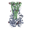
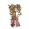
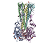
| Entry | Database: PDB / ID: 2ibx | |||||||||
|---|---|---|---|---|---|---|---|---|---|---|
| Title | Influenza virus (VN1194) H5 HA | |||||||||
 Components Components | (Hemagglutinin ) x 2 ) x 2 | |||||||||
 Keywords Keywords | VIRAL PROTEIN / Influenza / Haemagglutinin / H5N1 | |||||||||
| Function / homology |  Function and homology information Function and homology informationclathrin-dependent endocytosis of virus by host cell / membrane => GO:0016020 / host cell surface receptor binding / apical plasma membrane / fusion of virus membrane with host plasma membrane / fusion of virus membrane with host endosome membrane / viral envelope / virion attachment to host cell / host cell plasma membraneSimilarity search - Function | |||||||||
| Biological species |   Influenza A virus Influenza A virus | |||||||||
| Method | X-RAY DIFFRACTION / MOLECULAR REPLACEMENT / Resolution: 2.8 Å | |||||||||
 Authors Authors | Yamada, S. / Russell, R.J. / Gamblin, S.J. / Skehel, J.J. / Kawaoka, Y. | |||||||||
 Citation Citation | Journal: Nature / Year: 2006 Title: Haemagglutinin mutations responsible for the binding of H5N1 influenza A viruses to human-type receptors. Authors: Yamada, S. / Suzuki, Y. / Suzuki, T. / Le, M.Q. / Nidom, C.A. / Sakai-Tagawa, Y. / Muramoto, Y. / Ito, M. / Kiso, M. / Horimoto, T. / Shinya, K. / Sawada, T. / Kiso, M. / Usui, T. / Murata, ...Authors: Yamada, S. / Suzuki, Y. / Suzuki, T. / Le, M.Q. / Nidom, C.A. / Sakai-Tagawa, Y. / Muramoto, Y. / Ito, M. / Kiso, M. / Horimoto, T. / Shinya, K. / Sawada, T. / Kiso, M. / Usui, T. / Murata, T. / Lin, Y. / Hay, A. / Haire, L.F. / Stevens, D.J. / Russell, R.J. / Gamblin, S.J. / Skehel, J.J. / Kawaoka, Y. | |||||||||
| History |
|
- Structure visualization
| Structure viewer | Molecule: MolmilJmol/JSmol |
|---|
- Downloads & links
-Download
| PDBx/mmCIF format | 2ibx.cif.gz | 290.9 KB | Display | PDBx/mmCIF format |
|---|---|---|---|---|
| PDB format | pdb2ibx.ent.gz | 237.8 KB | Display | PDB format |
| PDBx/mmJSON format | 2ibx.json.gz | Tree view | PDBx/mmJSON format | |
| Others |  Other downloads Other downloads |
-Validation report
| Arichive directory | https://data.pdbj.org/pub/pdb/validation_reports/ib/2ibxftp://data.pdbj.org/pub/pdb/validation_reports/ib/2ibx | HTTPS FTP |
|---|
-Related structure data
| Related structure data | 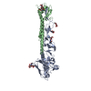 1jsmS S: Starting model for refinement |
|---|---|
| Similar structure data |


-Links
 PDBj
PDBj


- Assembly
| Deposited unit | 
| ||||||||
|---|---|---|---|---|---|---|---|---|---|
| 1 |
| ||||||||
| Unit cell |
|
-Components
| #1: Protein | / Fragment Mass: 38466.723 Da / Num. of mol.: 3 / Fragment: Residues 1-340 Source method: isolated from a genetically manipulated source Details: Polybasic cleavage site removed / Source: (gene. exp.) Influenza A virus / Genus: Influenzavirus A / Strain: H5N1 (Vn1194) / Gene: HA / References: UniProt: Q6DQ34, UniProt: Q45ZQ3*PLUS#2: Protein | / FragmentMass: 18390.303 Da / Num. of mol.: 3 / Fragment: Residues 347-506 Source method: isolated from a genetically manipulated source Details: Polybasic cleavage site removed. C-terminal part is bromelain cleaved Source: (gene. exp.) Influenza A virus / Genus: Influenzavirus A / Strain: H5N1 (Vn1194) / Gene: HA / References: UniProt: Q6DQ34, UniProt: Q66QC3*PLUS#3: Polysaccharide | / Mass: 424.401 Da / Num. of mol.: 3Source method: isolated from a genetically manipulated source #4: Sugar | ChemComp-NAG / N-Acetylglucosamine  Type: D-saccharide, beta linking / Mass: 221.208 Da / Num. of mol.: 6 Type: D-saccharide, beta linking / Mass: 221.208 Da / Num. of mol.: 6Source method: isolated from a genetically manipulated source Formula: C8H15NO6 |
|---|
-Experimental details
-Experiment
| Experiment | Method: X-RAY DIFFRACTION / Number of used crystals: 1 |
|---|
- Sample preparation
| Crystal | Density Matthews: 3.84 Å3/Da / Density % sol: 64.31 % |
|---|---|
| Crystal grow | Temperature: 293 K / pH: 6.5 Details: 15/4 EO/OH, Ammonium sulfate, Bis-tris, pH 6.50, VAPOR DIFFUSION, SITTING DROP, temperature 293K |
-Data collection
| Diffraction | Mean temperature: 100 K |
|---|---|
| Diffraction source | Source: ROTATING ANODE / Type: RIGAKU / Wavelength: 1.54178 |
| Detector | Type: RIGAKU RAXIS IV / Detector: IMAGE PLATE / Date: Feb 2, 2006 |
| Radiation | Monochromator: YALE MIRRORS / Protocol: SINGLE WAVELENGTH / Monochromatic (M) / Laue (L): M / Scattering type: x-ray |
| Radiation wavelength | Wavelength: 1.54178 Å / Relative weight: 1 |
| Reflection | Resolution: 2.8→30 Å / Num. obs: 48951 / % possible obs: 76.9 % / Observed criterion σ(I): 0 / Redundancy: 2.7 % / Rmerge(I) obs: 0.097 / Net I/σ(I): 8.7 |
| Reflection shell | Resolution: 2.8→2.93 Å / Rmerge(I) obs: 0.496 / Mean I/σ(I) obs: 1.8 / % possible all: 80.2 |
- Processing
| Software |
| ||||||||||||||||||||||||||||||||||||||||||||||||||||||||||||||||||||||||||||||||||||||||||||||||||||||||||||||||||||||||||||||||||||||||||||||||||||||||||||||||||||||||||
|---|---|---|---|---|---|---|---|---|---|---|---|---|---|---|---|---|---|---|---|---|---|---|---|---|---|---|---|---|---|---|---|---|---|---|---|---|---|---|---|---|---|---|---|---|---|---|---|---|---|---|---|---|---|---|---|---|---|---|---|---|---|---|---|---|---|---|---|---|---|---|---|---|---|---|---|---|---|---|---|---|---|---|---|---|---|---|---|---|---|---|---|---|---|---|---|---|---|---|---|---|---|---|---|---|---|---|---|---|---|---|---|---|---|---|---|---|---|---|---|---|---|---|---|---|---|---|---|---|---|---|---|---|---|---|---|---|---|---|---|---|---|---|---|---|---|---|---|---|---|---|---|---|---|---|---|---|---|---|---|---|---|---|---|---|---|---|---|---|---|---|---|
| Refinement | Method to determine structure: MOLECULAR REPLACEMENT Starting model: PDB ENTRY 1JSM Resolution: 2.8→30 Å / Cor.coef. Fo:Fc: 0.912 / Cor.coef. Fo:Fc free: 0.864 / SU B: 15.921 / SU ML: 0.316 / Cross valid method: THROUGHOUT / σ(F): 0 / ESU R Free: 0.474 / Stereochemistry target values: MAXIMUM LIKELIHOOD / Details: HYDROGENS HAVE BEEN ADDED IN THE RIDING POSITIONS
| ||||||||||||||||||||||||||||||||||||||||||||||||||||||||||||||||||||||||||||||||||||||||||||||||||||||||||||||||||||||||||||||||||||||||||||||||||||||||||||||||||||||||||
| Solvent computation | Ion probe radii: 0.8 Å / Shrinkage radii: 0.8 Å / VDW probe radii: 1.4 Å / Solvent model: MASK | ||||||||||||||||||||||||||||||||||||||||||||||||||||||||||||||||||||||||||||||||||||||||||||||||||||||||||||||||||||||||||||||||||||||||||||||||||||||||||||||||||||||||||
| Refinement step | Cycle: LAST / Resolution: 2.8→30 Å
| ||||||||||||||||||||||||||||||||||||||||||||||||||||||||||||||||||||||||||||||||||||||||||||||||||||||||||||||||||||||||||||||||||||||||||||||||||||||||||||||||||||||||||
| Refine LS restraints |
| ||||||||||||||||||||||||||||||||||||||||||||||||||||||||||||||||||||||||||||||||||||||||||||||||||||||||||||||||||||||||||||||||||||||||||||||||||||||||||||||||||||||||||
| LS refinement shell | Resolution: 2.8→2.87 Å / Total num. of bins used: 20
|
