 Movie
Movie Controller

[English] 日本語
 Yorodumi
- PDB-2ib8: Crystallographic and kinetic studies of human mitochondrial aceto... -
+ Open data

- Basic information
| Entry | Database: PDB / ID: 2ib8 | ||||||
|---|---|---|---|---|---|---|---|
| Title | Crystallographic and kinetic studies of human mitochondrial acetoacetyl-CoA thiolase (T2): the importance of potassium and chloride for its structure and function | ||||||
 Components Components | Acetyl-CoA acetyltransferase | ||||||
 Keywords Keywords |  TRANSFERASE / thiolase fold / potassium ion / chloride / beta-alpha-beta-alpha-beta-alpha-beta-beta topology / alpha-beta-alpha-beta-alpha layered structure TRANSFERASE / thiolase fold / potassium ion / chloride / beta-alpha-beta-alpha-beta-alpha-beta-beta topology / alpha-beta-alpha-beta-alpha layered structure | ||||||
| Function / homology |  Function and homology information Function and homology informationC-acetyltransferase activity / metanephric proximal convoluted tubule development / ketone body metabolic process / propionyl-CoA biosynthetic process / Utilization of Ketone Bodies / Synthesis of Ketone Bodies / cholesterol O-acyltransferase activity / ketone body catabolic process / acetyl-CoA catabolic process / isoleucine catabolic process ...C-acetyltransferase activity / metanephric proximal convoluted tubule development / ketone body metabolic process / propionyl-CoA biosynthetic process / Utilization of Ketone Bodies / Synthesis of Ketone Bodies / cholesterol O-acyltransferase activity / ketone body catabolic process / acetyl-CoA catabolic process / isoleucine catabolic process / acetyl-CoA C-acetyltransferase / acetyl-CoA C-acetyltransferase activity / coenzyme A binding / acetyl-CoA biosynthetic process / Branched-chain amino acid catabolism / coenzyme A metabolic process / coenzyme A biosynthetic process / fatty acid beta-oxidation / response to starvation / potassium ion binding / Mitochondrial protein degradation / adipose tissue development / liver development / response to hormone / response to organic cyclic compound / mitochondrial matrix / enzyme binding / endoplasmic reticulum / mitochondrion / extracellular exosome / identical protein bindingSimilarity search - Function | ||||||
| Biological species |  Homo sapiens (human) Homo sapiens (human) | ||||||
| Method | X-RAY DIFFRACTION / SYNCHROTRON / MOLECULAR REPLACEMENT / Resolution: 1.85 Å | ||||||
 Authors Authors | Haapalainen, A.M. / Wierenga, R.K. | ||||||
 Citation Citation | Journal: Biochemistry / Year: 2007 Title: Crystallographic and Kinetic Studies of Human Mitochondrial Acetoacetyl-CoA Thiolase: The Importance of Potassium and Chloride Ions for Its Structure and Function Authors: Haapalainen, A.M. / Merilainen, G. / Pirila, P.L. / Kondo, N. / Fukao, T. / Wierenga, R.K. | ||||||
| History |
|
- Structure visualization
| Structure viewer | Molecule: MolmilJmol/JSmol |
|---|
- Downloads & links
-Download
| PDBx/mmCIF format | 2ib8.cif.gz | 320.6 KB | Display | PDBx/mmCIF format |
|---|---|---|---|---|
| PDB format | pdb2ib8.ent.gz | 258.1 KB | Display | PDB format |
| PDBx/mmJSON format | 2ib8.json.gz | Tree view | PDBx/mmJSON format | |
| Others |  Other downloads Other downloads |
-Validation report
| Arichive directory | https://data.pdbj.org/pub/pdb/validation_reports/ib/2ib8ftp://data.pdbj.org/pub/pdb/validation_reports/ib/2ib8 | HTTPS FTP |
|---|
-Related structure data
| Related structure data |  2ib7C  2ib9C  2ibuC  2ibwC 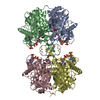 2ibyC  1wl4S C: citing same article ( S: Starting model for refinement |
|---|---|
| Similar structure data |


-Links
 PDBj
PDBj

- Assembly
| Deposited unit | 
| ||||||||
|---|---|---|---|---|---|---|---|---|---|
| 1 |
| ||||||||
| Unit cell |
| ||||||||
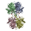
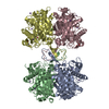
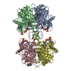
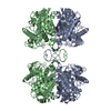
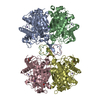
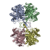
| Details | The asymmetric unit consists of one biological unit, the homotetramer |
-Components
-Protein , 1 types, 4 molecules ABCD
| #1: Protein | Mass: 41533.047 Da / Num. of mol.: 4 / Mutation: V34A Source method: isolated from a genetically manipulated source Source: (gene. exp.) Homo sapiens (human) / Tissue: liver / Gene: ACAT1 / Plasmid: pET3D / Production host:  Escherichia coli (E. coli) / Strain (production host): BL21 (DE3) pLysS / References: UniProt: P24752, acetyl-CoA C-acetyltransferase Escherichia coli (E. coli) / Strain (production host): BL21 (DE3) pLysS / References: UniProt: P24752, acetyl-CoA C-acetyltransferase |
|---|
-Non-polymers , 5 types, 1050 molecules 


| #2: Chemical | ChemComp-CL / Chloride Mass: 35.453 Da / Num. of mol.: 4 / Source method: obtained synthetically / Formula: Cl Mass: 35.453 Da / Num. of mol.: 4 / Source method: obtained synthetically / Formula: Cl#3: Chemical | ChemComp-K /  Mass: 39.098 Da / Num. of mol.: 4 / Source method: obtained synthetically / Formula: K Mass: 39.098 Da / Num. of mol.: 4 / Source method: obtained synthetically / Formula: K#4: Chemical | ChemComp-MES / MES (buffer) Mass: 195.237 Da / Num. of mol.: 4 / Source method: obtained synthetically / Formula: C6H13NO4S / Comment: pH buffer*YM Mass: 195.237 Da / Num. of mol.: 4 / Source method: obtained synthetically / Formula: C6H13NO4S / Comment: pH buffer*YM#5: Chemical | ChemComp-GOL / Glycerol Mass: 92.094 Da / Num. of mol.: 5 / Source method: obtained synthetically / Formula: C3H8O3 Mass: 92.094 Da / Num. of mol.: 5 / Source method: obtained synthetically / Formula: C3H8O3#6: Water | ChemComp-HOH / | WaterMass: 18.015 Da / Num. of mol.: 1033 / Source method: isolated from a natural source / Formula: H2O |
|---|
-Experimental details
-Experiment
| Experiment | Method: X-RAY DIFFRACTION / Number of used crystals: 1 |
|---|
- Sample preparation
| Crystal | Density Matthews: 2.43 Å3/Da / Density % sol: 49.51 % |
|---|---|
| Crystal grow | Temperature: 277 K / Method: vapor diffusion, hanging drop / pH: 6.5 Details: 18% PEG 5000 monomethylether, 0.1M MES, pH 6.5, VAPOR DIFFUSION, HANGING DROP, temperature 277.0K |
-Data collection
| Diffraction | Mean temperature: 100 K |
|---|---|
| Diffraction source | Source: SYNCHROTRON / Site: MAX II  / Beamline: I711 / Wavelength: 1.141 Å / Beamline: I711 / Wavelength: 1.141 Å |
| Detector | Type: MAR CCD 165 mm / Detector: CCD / Date: Dec 17, 2005 / Details: mirrors |
| Radiation | Monochromator: Single asymmetrically cut Si(111) crystal with horizontal diffraction plane Protocol: SINGLE WAVELENGTH / Monochromatic (M) / Laue (L): M / Scattering type: x-ray |
| Radiation wavelength | Wavelength: 1.141 Å / Relative weight: 1 |
| Reflection | Resolution: 1.85→47.25 Å / Num. all: 135350 / Num. obs: 135257 / % possible obs: 99.9 % / Observed criterion σ(F): 0 / Observed criterion σ(I): 0 / Redundancy: 4.2 % / Biso Wilson estimate: 24 Å2 / Rmerge(I) obs: 0.061 / Rsym value: 0.061 / Net I/σ(I): 16.9 |
| Reflection shell | Resolution: 1.85→1.95 Å / Redundancy: 4.1 % / Rmerge(I) obs: 0.301 / Mean I/σ(I) obs: 4.6 / Num. unique all: 19653 / Rsym value: 0.301 / % possible all: 100 |
- Processing
| Software |
| |||||||||||||||||||||||||||||||||||||||||||||||||||||||||||||||||||||||||||||||||||||||||||||||
|---|---|---|---|---|---|---|---|---|---|---|---|---|---|---|---|---|---|---|---|---|---|---|---|---|---|---|---|---|---|---|---|---|---|---|---|---|---|---|---|---|---|---|---|---|---|---|---|---|---|---|---|---|---|---|---|---|---|---|---|---|---|---|---|---|---|---|---|---|---|---|---|---|---|---|---|---|---|---|---|---|---|---|---|---|---|---|---|---|---|---|---|---|---|---|---|---|
| Refinement | Method to determine structure: MOLECULAR REPLACEMENT Starting model: PDB entry 1WL4 Resolution: 1.85→47.25 Å / Cor.coef. Fo:Fc: 0.961 / Cor.coef. Fo:Fc free: 0.934 / SU B: 2.646 / SU ML: 0.081 / Cross valid method: THROUGHOUT / σ(F): 0 / σ(I): 0 / ESU R: 0.124 / ESU R Free: 0.122 / Stereochemistry target values: MAXIMUM LIKELIHOOD
| |||||||||||||||||||||||||||||||||||||||||||||||||||||||||||||||||||||||||||||||||||||||||||||||
| Solvent computation | Ion probe radii: 0.8 Å / Shrinkage radii: 0.8 Å / VDW probe radii: 1.2 Å / Solvent model: BABINET MODEL WITH MASK | |||||||||||||||||||||||||||||||||||||||||||||||||||||||||||||||||||||||||||||||||||||||||||||||
| Displacement parameters | Biso mean: 24.195 Å2
| |||||||||||||||||||||||||||||||||||||||||||||||||||||||||||||||||||||||||||||||||||||||||||||||
| Refinement step | Cycle: LAST / Resolution: 1.85→47.25 Å
| |||||||||||||||||||||||||||||||||||||||||||||||||||||||||||||||||||||||||||||||||||||||||||||||
| Refine LS restraints |
| |||||||||||||||||||||||||||||||||||||||||||||||||||||||||||||||||||||||||||||||||||||||||||||||
| LS refinement shell | Resolution: 1.85→1.915 Å / Total num. of bins used: 15
|
