 Movie
Movie Controller


+ Open data

- Basic information
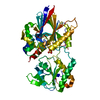
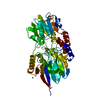
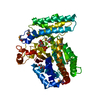
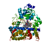
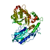
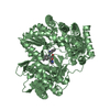
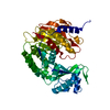
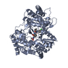
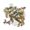
| Entry | Database: PDB / ID: 2i7x | ||||||
|---|---|---|---|---|---|---|---|
| Title | Structure of Yeast CPSF-100 (Ydh1p) | ||||||
 Components Components | Protein CFT2 | ||||||
 Keywords Keywords |  RNA BINDING PROTEIN / PROTEIN BINDING / polyadenylation / metallo-B-lactamase / pre-mRNA processing / Artemis / V(D)J recombination / double-strand break repair RNA BINDING PROTEIN / PROTEIN BINDING / polyadenylation / metallo-B-lactamase / pre-mRNA processing / Artemis / V(D)J recombination / double-strand break repair | ||||||
| Function / homology |  Function and homology information Function and homology informationProcessing of Intronless Pre-mRNAs / termination of RNA polymerase II transcription, poly(A)-coupled / mRNA cleavage and polyadenylation specificity factor complex / : / mRNA processing / RNA binding / nucleusSimilarity search - Function | ||||||
| Biological species |  Saccharomyces cerevisiae (brewer's yeast) Saccharomyces cerevisiae (brewer's yeast) | ||||||
| Method | X-RAY DIFFRACTION / SYNCHROTRON / SAD / Resolution: 2.5 Å | ||||||
 Authors Authors | Mandel, C.R. / Zhang, H. / Gebauer, D. / Tong, L. | ||||||
 Citation Citation | Journal: Nature / Year: 2006 Title: Polyadenylation factor CPSF-73 is the pre-mRNA 3'-end-processing endonuclease. Authors: Mandel, C.R. / Kaneko, S. / Zhang, H. / Gebauer, D. / Vethantham, V. / Manley, J.L. / Tong, L. | ||||||
| History |
|
- Structure visualization
| Structure viewer | Molecule: MolmilJmol/JSmol |
|---|
- Downloads & links
-Download
| PDBx/mmCIF format | 2i7x.cif.gz | 114.4 KB | Display | PDBx/mmCIF format |
|---|---|---|---|---|
| PDB format | pdb2i7x.ent.gz | 87.4 KB | Display | PDB format |
| PDBx/mmJSON format | 2i7x.json.gz | Tree view | PDBx/mmJSON format | |
| Others |  Other downloads Other downloads |
-Validation report
| Arichive directory | https://data.pdbj.org/pub/pdb/validation_reports/i7/2i7xftp://data.pdbj.org/pub/pdb/validation_reports/i7/2i7x | HTTPS FTP |
|---|
-Related structure data


-Links
 PDBj
PDBj


- Assembly
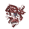
| Deposited unit | 
| ||||||||
|---|---|---|---|---|---|---|---|---|---|
| 1 |
| ||||||||
| Unit cell |
|
-Components
| #1: Protein | Mass: 80677.695 Da / Num. of mol.: 1 Source method: isolated from a genetically manipulated source Source: (gene. exp.) Saccharomyces cerevisiae (brewer's yeast)Gene: CFT2, YDH1 / Plasmid: pET-28a / Production host:  Escherichia coli (E. coli) / References: UniProt: Q12102 Escherichia coli (E. coli) / References: UniProt: Q12102 |
|---|---|
| #2: Water | ChemComp-HOH / Water Mass: 18.015 Da / Num. of mol.: 148 / Source method: isolated from a natural source / Formula: H2O Mass: 18.015 Da / Num. of mol.: 148 / Source method: isolated from a natural source / Formula: H2O |
-Experimental details
-Experiment
| Experiment | Method: X-RAY DIFFRACTION / Number of used crystals: 1 |
|---|
- Sample preparation
| Crystal | Density Matthews: 1.82 Å3/Da / Density % sol: 32.59 % |
|---|---|
| Crystal grow | Temperature: 277 K / Method: vapor diffusion, hanging drop / pH: 8.5 Details: 20% PEG 3350, 0.2M ammonium citrate, pH 8.5, VAPOR DIFFUSION, HANGING DROP, temperature 277K |
-Data collection
| Diffraction | Mean temperature: 100 K |
|---|---|
| Diffraction source | Source: SYNCHROTRON / Site: NSLS  / Beamline: X4A / Wavelength: 0.9798 / Beamline: X4A / Wavelength: 0.9798 |
| Detector | Type: ADSC QUANTUM 4 / Detector: CCD / Details: MIRRORS |
| Radiation | Monochromator: SI(111) / Protocol: SINGLE WAVELENGTH / Monochromatic (M) / Laue (L): M / Scattering type: x-ray |
| Radiation wavelength | Wavelength: 0.9798 Å / Relative weight: 1 |
| Reflection | Resolution: 2.5→30 Å / Num. obs: 20806 / % possible obs: 99.9 % / Redundancy: 11.3 % / Biso Wilson estimate: 37.1 Å2 / Rmerge(I) obs: 0.087 / Net I/σ(I): 24.4674 |
| Reflection shell | Resolution: 2.5→2.59 Å / Redundancy: 10.4 % / Rmerge(I) obs: 0.408 / Mean I/σ(I) obs: 5.644 / % possible all: 100 |
- Processing
| Software |
| ||||||||||||||||||||||||||||||||||||
|---|---|---|---|---|---|---|---|---|---|---|---|---|---|---|---|---|---|---|---|---|---|---|---|---|---|---|---|---|---|---|---|---|---|---|---|---|---|
| Refinement | Method to determine structure: SAD / Resolution: 2.5→28.18 Å / Rfactor Rfree error: 0.007 / Data cutoff high absF: 459366.6 / Data cutoff low absF: 0 / Isotropic thermal model: RESTRAINED / Cross valid method: THROUGHOUT / σ(F): 2
| ||||||||||||||||||||||||||||||||||||
| Solvent computation | Solvent model: FLAT MODEL / Bsol: 31.7429 Å2 / ksol: 0.301021 e/Å3 | ||||||||||||||||||||||||||||||||||||
| Displacement parameters | Biso mean: 50.8 Å2
| ||||||||||||||||||||||||||||||||||||
| Refine analyze |
| ||||||||||||||||||||||||||||||||||||
| Refinement step | Cycle: LAST / Resolution: 2.5→28.18 Å
| ||||||||||||||||||||||||||||||||||||
| Refine LS restraints |
| ||||||||||||||||||||||||||||||||||||
| LS refinement shell | Resolution: 2.5→2.59 Å / Rfactor Rfree error: 0.032 / Total num. of bins used: 10
| ||||||||||||||||||||||||||||||||||||
| Xplor file |
|
