 Movie
Movie Controller


+ Open data

- Basic information
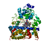
| Entry | Database: PDB / ID: 4tx2 | |||||||||
|---|---|---|---|---|---|---|---|---|---|---|
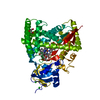
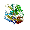
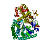
| Title | Crystal structure of the X-domain from teicoplanin biosynthesis | |||||||||
 Components Components | Non-ribosomal peptide synthetase | |||||||||
 Keywords Keywords |  PROTEIN BINDING / non-ribosomal peptide synthetase / condensation type domain / teicoplanin biosynthesis / oxygenase complex PROTEIN BINDING / non-ribosomal peptide synthetase / condensation type domain / teicoplanin biosynthesis / oxygenase complex | |||||||||
| Function / homology |  Function and homology information Function and homology informationamide biosynthetic process / : / organonitrogen compound biosynthetic process / secondary metabolite biosynthetic process / lipid biosynthetic process / phosphopantetheine binding / catalytic activitySimilarity search - Function | |||||||||
| Biological species |  Actinoplanes teichomyceticus (bacteria) Actinoplanes teichomyceticus (bacteria) | |||||||||
| Method | X-RAY DIFFRACTION / SYNCHROTRON / MOLECULAR REPLACEMENT / Resolution: 2.9 Å | |||||||||
 Authors Authors | Peschke, M. / Haslinger, K. / Cryle, M.J. | |||||||||
| Funding support |  Germany, 1items Germany, 1items
| |||||||||
 Citation Citation | Journal: Nature / Year: 2015 Title: X-domain of peptide synthetases recruits oxygenases crucial for glycopeptide biosynthesis. Authors: Haslinger, K. / Peschke, M. / Brieke, C. / Maximowitsch, E. / Cryle, M.J. | |||||||||
| History |
|
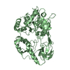
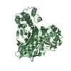
- Structure visualization
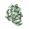
| Structure viewer | Molecule: MolmilJmol/JSmol |
|---|
- Downloads & links
-Download
| PDBx/mmCIF format | 4tx2.cif.gz | 102 KB | Display | PDBx/mmCIF format |
|---|---|---|---|---|
| PDB format | pdb4tx2.ent.gz | 76 KB | Display | PDB format |
| PDBx/mmJSON format | 4tx2.json.gz | Tree view | PDBx/mmJSON format | |
| Others |  Other downloads Other downloads |
-Validation report
| Arichive directory | https://data.pdbj.org/pub/pdb/validation_reports/tx/4tx2ftp://data.pdbj.org/pub/pdb/validation_reports/tx/4tx2 | HTTPS FTP |
|---|
-Related structure data
| Related structure data |  4tx3C 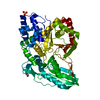 2jgpS S: Starting model for refinement C: citing same article ( |
|---|---|
| Similar structure data |
-Links
 PDBj
PDBj


- Assembly
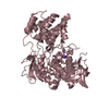
| Deposited unit | 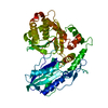
| ||||||||
|---|---|---|---|---|---|---|---|---|---|
| 1 |
| ||||||||
| Unit cell |
|
-Components
| #1: Protein | Mass: 53435.977 Da / Num. of mol.: 1 / Fragment: UNP residues 1047-1511 Source method: isolated from a genetically manipulated source Source: (gene. exp.) Actinoplanes teichomyceticus (bacteria)Gene: tcp12 / Plasmid: pET24d / Details (production host): GB1 fusion protein / Production host: Escherichia coli BL21(DE3) (bacteria) / References: UniProt: Q70AZ6 |
|---|---|
| #2: Water | ChemComp-HOH / Water Mass: 18.015 Da / Num. of mol.: 18 / Source method: isolated from a natural source / Formula: H2O Mass: 18.015 Da / Num. of mol.: 18 / Source method: isolated from a natural source / Formula: H2O |
-Experimental details
-Experiment
| Experiment | Method: X-RAY DIFFRACTION / Number of used crystals: 1 |
|---|
- Sample preparation
| Crystal | Density Matthews: 2.68 Å3/Da / Density % sol: 54.04 % |
|---|---|
| Crystal grow | Temperature: 293 K / Method: vapor diffusion, hanging drop / pH: 6.5 / Details: 100 mM Bis-Tris, 200 mM (NH4)2SO4, 20% PEG 3350 |
-Data collection
| Diffraction | Mean temperature: 100 K | ||||||||||||||||||||||||||||||||||||||||||||||||||||||||||||||||||||||||||||||||||||||||||||||||||||||||||||||||||||||||||||||||||||||||||||||||||||||||||||||||||||||||||||||||||||
|---|---|---|---|---|---|---|---|---|---|---|---|---|---|---|---|---|---|---|---|---|---|---|---|---|---|---|---|---|---|---|---|---|---|---|---|---|---|---|---|---|---|---|---|---|---|---|---|---|---|---|---|---|---|---|---|---|---|---|---|---|---|---|---|---|---|---|---|---|---|---|---|---|---|---|---|---|---|---|---|---|---|---|---|---|---|---|---|---|---|---|---|---|---|---|---|---|---|---|---|---|---|---|---|---|---|---|---|---|---|---|---|---|---|---|---|---|---|---|---|---|---|---|---|---|---|---|---|---|---|---|---|---|---|---|---|---|---|---|---|---|---|---|---|---|---|---|---|---|---|---|---|---|---|---|---|---|---|---|---|---|---|---|---|---|---|---|---|---|---|---|---|---|---|---|---|---|---|---|---|---|---|
| Diffraction source | Source: SYNCHROTRON / Site: SLS  / Beamline: X10SA / Wavelength: 0.9797 Å / Beamline: X10SA / Wavelength: 0.9797 Å | ||||||||||||||||||||||||||||||||||||||||||||||||||||||||||||||||||||||||||||||||||||||||||||||||||||||||||||||||||||||||||||||||||||||||||||||||||||||||||||||||||||||||||||||||||||
| Detector | Type: PSI PILATUS 6M / Detector: PIXEL / Date: Feb 24, 2014 | ||||||||||||||||||||||||||||||||||||||||||||||||||||||||||||||||||||||||||||||||||||||||||||||||||||||||||||||||||||||||||||||||||||||||||||||||||||||||||||||||||||||||||||||||||||
| Radiation | Monochromator: Si(111) / Protocol: SINGLE WAVELENGTH / Monochromatic (M) / Laue (L): M / Scattering type: x-ray | ||||||||||||||||||||||||||||||||||||||||||||||||||||||||||||||||||||||||||||||||||||||||||||||||||||||||||||||||||||||||||||||||||||||||||||||||||||||||||||||||||||||||||||||||||||
| Radiation wavelength | Wavelength: 0.9797 Å / Relative weight: 1 | ||||||||||||||||||||||||||||||||||||||||||||||||||||||||||||||||||||||||||||||||||||||||||||||||||||||||||||||||||||||||||||||||||||||||||||||||||||||||||||||||||||||||||||||||||||
| Reflection | Resolution: 2.9→48 Å / Num. obs: 12768 / % possible obs: 99.9 % / Observed criterion σ(I): -3 / Redundancy: 20.5 % / Biso Wilson estimate: 42.4 Å2 / Rmerge F obs: 0.999 / Rmerge(I) obs: 0.119 / Rrim(I) all: 0.122 / Χ2: 0.914 / Net I/σ(I): 24.31 / Num. measured all: 261201 | ||||||||||||||||||||||||||||||||||||||||||||||||||||||||||||||||||||||||||||||||||||||||||||||||||||||||||||||||||||||||||||||||||||||||||||||||||||||||||||||||||||||||||||||||||||
| Reflection shell | Diffraction-ID: 1 / Rejects: 0
|
- Processing
| Software |
| ||||||||||||||||||||||||||||||||||||
|---|---|---|---|---|---|---|---|---|---|---|---|---|---|---|---|---|---|---|---|---|---|---|---|---|---|---|---|---|---|---|---|---|---|---|---|---|---|
| Refinement | Method to determine structure: MOLECULAR REPLACEMENT Starting model: 2JGP Resolution: 2.9→44.186 Å / SU ML: 0.44 / Cross valid method: THROUGHOUT / σ(F): 1.37 / Phase error: 25.25 / Stereochemistry target values: ML
| ||||||||||||||||||||||||||||||||||||
| Solvent computation | Shrinkage radii: 0.9 Å / VDW probe radii: 1.11 Å / Solvent model: FLAT BULK SOLVENT MODEL | ||||||||||||||||||||||||||||||||||||
| Displacement parameters | Biso max: 103.88 Å2 / Biso mean: 30.6939 Å2 / Biso min: 3.42 Å2 | ||||||||||||||||||||||||||||||||||||
| Refinement step | Cycle: final / Resolution: 2.9→44.186 Å
| ||||||||||||||||||||||||||||||||||||
| Refine LS restraints |
| ||||||||||||||||||||||||||||||||||||
| LS refinement shell | Refine-ID: X-RAY DIFFRACTION / Total num. of bins used: 5 / % reflection obs: 100 %
|
