 Movie
Movie Controller

[English] 日本語
 Yorodumi
- PDB-2i71: Crystal structure of a Conserved Protein of Unknown Function from... -
+ Open data

- Basic information
| Entry | Database: PDB / ID: 2i71 | ||||||
|---|---|---|---|---|---|---|---|
| Title | Crystal structure of a Conserved Protein of Unknown Function from Sulfolobus solfataricus P2 | ||||||
 Components Components | Hypothetical protein | ||||||
 Keywords Keywords | STRUCTURAL GENOMICS / UNKNOWN FUNCTION / APC6294 / Conserved hypothetical protein / Sulfolobus solfataricus P2 / PSI-2 / Protein structure initiative / Midwest center for structural genomics / MCSG | ||||||
| Function / homology |  Function and homology informationendonuclease activity / defense response to virus / Hydrolases; Acting on ester bonds Function and homology informationendonuclease activity / defense response to virus / Hydrolases; Acting on ester bondsSimilarity search - Function | ||||||
| Biological species |   Sulfolobus solfataricus (archaea) Sulfolobus solfataricus (archaea) | ||||||
| Method | X-RAY DIFFRACTION / SYNCHROTRON / SAD / Resolution: 1.7 Å | ||||||
 Authors Authors | Tan, K. / Skarina, T. / Onopriyenko, O. / Savchenko, A. / Edwards, A. / Joachimiak, A. / Midwest Center for Structural Genomics (MCSG) | ||||||
 Citation Citation | Journal: To be Published Title: The crystal structure of a Conserved hypothetical protein from Sulfolobus solfataricus P2 Authors: Tan, K. / Skarina, T. / Onopriyenko, O. / Savchenko, A. / Edwards, A. / Joachimiak, A. | ||||||
| History |
| ||||||
| Remark 300 | THIS ENTRY CONTAINS THE CRYSTALLOGRAPHIC ASYMMETRIC UNIT WHICH CONSISTS OF 2 CHAINS. THE AUTHOR ... THIS ENTRY CONTAINS THE CRYSTALLOGRAPHIC ASYMMETRIC UNIT WHICH CONSISTS OF 2 CHAINS. THE AUTHOR STATES THAT THE BIOLOGICAL UNIT OF THIS PROTEIN IS UNKNOWN. |
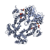
- Structure visualization
| Structure viewer | Molecule: MolmilJmol/JSmol |
|---|
- Downloads & links
-Download
| PDBx/mmCIF format | 2i71.cif.gz | 173.6 KB | Display | PDBx/mmCIF format |
|---|---|---|---|---|
| PDB format | pdb2i71.ent.gz | 142.3 KB | Display | PDB format |
| PDBx/mmJSON format | 2i71.json.gz | Tree view | PDBx/mmJSON format | |
| Others |  Other downloads Other downloads |
-Validation report
| Arichive directory | https://data.pdbj.org/pub/pdb/validation_reports/i7/2i71ftp://data.pdbj.org/pub/pdb/validation_reports/i7/2i71 | HTTPS FTP |
|---|
-Related structure data
| Similar structure data | |
|---|---|
| Other databases |


-Links
 PDBj
PDBj- Assembly
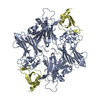
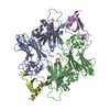
| Deposited unit | 
| ||||||||
|---|---|---|---|---|---|---|---|---|---|
| 1 |
| ||||||||
| 2 | 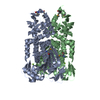
| ||||||||
| Unit cell |
|
-Components
| #1: Protein | Mass: 45843.992 Da / Num. of mol.: 2 Source method: isolated from a genetically manipulated source Source: (gene. exp.) Sulfolobus solfataricus (archaea) / Strain: P2 / Gene: SSO1389 / Plasmid: p15Tv lic / Species (production host): Escherichia coli / Production host:  Escherichia coli BL21 (bacteria) / Strain (production host): BL21 / References: UniProt: Q97YD5 Escherichia coli BL21 (bacteria) / Strain (production host): BL21 / References: UniProt: Q97YD5#2: Chemical |   Mass: 24.305 Da / Num. of mol.: 2 / Source method: obtained synthetically / Formula: Mg Mass: 24.305 Da / Num. of mol.: 2 / Source method: obtained synthetically / Formula: Mg#3: Water | ChemComp-HOH / | Water Mass: 18.015 Da / Num. of mol.: 786 / Source method: isolated from a natural source / Formula: H2O Mass: 18.015 Da / Num. of mol.: 786 / Source method: isolated from a natural source / Formula: H2O |
|---|
-Experimental details
-Experiment
| Experiment | Method: X-RAY DIFFRACTION / Number of used crystals: 1 |
|---|
- Sample preparation
| Crystal | Density Matthews: 1.98 Å3/Da / Density % sol: 37.9 % |
|---|---|
| Crystal grow | Temperature: 283 K / Method: vapor diffusion, sitting drop / pH: 5.5 Details: 25% PEG3350, 0.2M MgCl2, 0.1M BisTris, 2% Tacsimate, pH 5.5, VAPOR DIFFUSION, SITTING DROP, temperature 283K |
-Data collection
| Diffraction | Mean temperature: 100 K |
|---|---|
| Diffraction source | Source: SYNCHROTRON / Site: APS  / Beamline: 19-BM / Wavelength: 0.97915 Å / Beamline: 19-BM / Wavelength: 0.97915 Å |
| Detector | Type: SBC-3 / Detector: CCD / Date: Jun 21, 2006 / Details: mirror |
| Radiation | Monochromator: Si 111 Channel / Protocol: SINGLE WAVELENGTH / Monochromatic (M) / Laue (L): M / Scattering type: x-ray |
| Radiation wavelength | Wavelength: 0.97915 Å / Relative weight: 1 |
| Reflection | Resolution: 1.7→27.92 Å / Num. all: 80005 / Num. obs: 80005 / % possible obs: 99.8 % / Observed criterion σ(F): 0 / Observed criterion σ(I): 0 / Redundancy: 7 % / Biso Wilson estimate: 18.25 Å2 / Rmerge(I) obs: 0.086 / Net I/σ(I): 28.98 |
| Reflection shell | Resolution: 1.7→1.74 Å / Redundancy: 6.7 % / Rmerge(I) obs: 0.492 / Mean I/σ(I) obs: 3.7 / Num. unique all: 5265 / % possible all: 100 |
- Processing
| Software |
| ||||||||||||||||||||||||||||||||||||||||||||||||||||||||||||||||||||||||||||||||||||||||||||||||||||||||||||||||||||||||||||||||||||||||||||||||||||||||||||||||||||||||||
|---|---|---|---|---|---|---|---|---|---|---|---|---|---|---|---|---|---|---|---|---|---|---|---|---|---|---|---|---|---|---|---|---|---|---|---|---|---|---|---|---|---|---|---|---|---|---|---|---|---|---|---|---|---|---|---|---|---|---|---|---|---|---|---|---|---|---|---|---|---|---|---|---|---|---|---|---|---|---|---|---|---|---|---|---|---|---|---|---|---|---|---|---|---|---|---|---|---|---|---|---|---|---|---|---|---|---|---|---|---|---|---|---|---|---|---|---|---|---|---|---|---|---|---|---|---|---|---|---|---|---|---|---|---|---|---|---|---|---|---|---|---|---|---|---|---|---|---|---|---|---|---|---|---|---|---|---|---|---|---|---|---|---|---|---|---|---|---|---|---|---|---|
| Refinement | Method to determine structure: SAD / Resolution: 1.7→27.92 Å / Cor.coef. Fo:Fc: 0.958 / Cor.coef. Fo:Fc free: 0.934 / SU B: 2.165 / SU ML: 0.073 / Cross valid method: THROUGHOUT / σ(F): 0 / ESU R: 0.117 / ESU R Free: 0.118 / Stereochemistry target values: MAXIMUM LIKELIHOOD / Details: HYDROGENS HAVE BEEN ADDED IN THE RIDING POSITIONS
| ||||||||||||||||||||||||||||||||||||||||||||||||||||||||||||||||||||||||||||||||||||||||||||||||||||||||||||||||||||||||||||||||||||||||||||||||||||||||||||||||||||||||||
| Solvent computation | Ion probe radii: 0.8 Å / Shrinkage radii: 0.8 Å / VDW probe radii: 1.2 Å / Solvent model: MASK | ||||||||||||||||||||||||||||||||||||||||||||||||||||||||||||||||||||||||||||||||||||||||||||||||||||||||||||||||||||||||||||||||||||||||||||||||||||||||||||||||||||||||||
| Displacement parameters | Biso mean: 20.114 Å2
| ||||||||||||||||||||||||||||||||||||||||||||||||||||||||||||||||||||||||||||||||||||||||||||||||||||||||||||||||||||||||||||||||||||||||||||||||||||||||||||||||||||||||||
| Refinement step | Cycle: LAST / Resolution: 1.7→27.92 Å
| ||||||||||||||||||||||||||||||||||||||||||||||||||||||||||||||||||||||||||||||||||||||||||||||||||||||||||||||||||||||||||||||||||||||||||||||||||||||||||||||||||||||||||
| Refine LS restraints |
| ||||||||||||||||||||||||||||||||||||||||||||||||||||||||||||||||||||||||||||||||||||||||||||||||||||||||||||||||||||||||||||||||||||||||||||||||||||||||||||||||||||||||||
| LS refinement shell | Resolution: 1.7→1.745 Å / Total num. of bins used: 20
|
