 Movie
Movie Controller

[English] 日本語
 Yorodumi
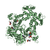
- PDB-2nz1: Viral Chemokine Binding Protein M3 From Murine Gammaherpesvirus68... -
+ Open data

- Basic information
| Entry | Database: PDB / ID: 2nz1 | ||||||
|---|---|---|---|---|---|---|---|
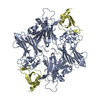
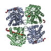
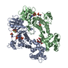
| Title | Viral Chemokine Binding Protein M3 From Murine Gammaherpesvirus68 In Complex With The CC-Chemokine CCL2/MCP-1 | ||||||
 Components Components |
| ||||||
 Keywords Keywords |  VIRAL PROTEIN/CYTOKINE / Viral Decoy Receptor / Chemokine / Protein-Protein Complex / VIRAL PROTEIN-CYTOKINE COMPLEX VIRAL PROTEIN/CYTOKINE / Viral Decoy Receptor / Chemokine / Protein-Protein Complex / VIRAL PROTEIN-CYTOKINE COMPLEX | ||||||
| Function / homology |  Function and homology information Function and homology informationhelper T cell extravasation / CCR2 chemokine receptor binding / negative regulation of natural killer cell chemotaxis / positive regulation of NMDA glutamate receptor activity / chemokine binding / negative regulation of glial cell apoptotic process / astrocyte cell migration / ATF4 activates genes in response to endoplasmic reticulum stress / positive regulation of apoptotic cell clearance / CCR chemokine receptor binding ...helper T cell extravasation / CCR2 chemokine receptor binding / negative regulation of natural killer cell chemotaxis / positive regulation of NMDA glutamate receptor activity / chemokine binding / negative regulation of glial cell apoptotic process / astrocyte cell migration / ATF4 activates genes in response to endoplasmic reticulum stress / positive regulation of apoptotic cell clearance / CCR chemokine receptor binding / lymphocyte chemotaxis / cellular homeostasis / NFE2L2 regulating inflammation associated genes / eosinophil chemotaxis / positive regulation of endothelial cell apoptotic process / cellular response to fibroblast growth factor stimulus / chemokine-mediated signaling pathway / Chemokine receptors bind chemokines / chemokine activity / negative regulation of vascular endothelial cell proliferation / negative regulation of G1/S transition of mitotic cell cycle / positive regulation of calcium ion import / macrophage chemotaxis / positive regulation of nitric-oxide synthase biosynthetic process / Interleukin-10 signaling / monocyte chemotaxis / cell surface receptor signaling pathway via JAK-STAT / G protein-coupled receptor signaling pathway, coupled to cyclic nucleotide second messenger / humoral immune response / cellular response to interleukin-1 / sensory perception of pain / cytoskeleton organization / viral genome replication / positive regulation of synaptic transmission, glutamatergic / neutrophil chemotaxis / animal organ morphogenesis / response to bacterium / cytokine-mediated signaling pathway / cellular response to type II interferon / chemotaxis / positive regulation of T cell activation / cellular response to tumor necrosis factor / regulation of cell shape / Interleukin-4 and Interleukin-13 signaling / angiogenesis / cellular response to lipopolysaccharide / negative regulation of neuron apoptotic process / cell surface receptor signaling pathway / positive regulation of ERK1 and ERK2 cascade / cell adhesion / protein kinase activity / inflammatory response / G protein-coupled receptor signaling pathway / protein phosphorylation / signaling receptor binding / signal transduction / extracellular space / extracellular regionSimilarity search - Function | ||||||
| Biological species |  Murid herpesvirus 4 (Murine herpesvirus 68) Murid herpesvirus 4 (Murine herpesvirus 68) Homo sapiens (human) Homo sapiens (human) | ||||||
| Method | X-RAY DIFFRACTION / SYNCHROTRON / MOLECULAR REPLACEMENT / Resolution: 2.5 Å | ||||||
 Authors Authors | Alexander-Brett, J.M. / Fremont, D.H. | ||||||
 Citation Citation | Journal: J.Exp.Med. / Year: 2007 Title: Dual GPCR and GAG mimicry by the M3 chemokine decoy receptor. Authors: Alexander-Brett, J.M. / Fremont, D.H. | ||||||
| History |
|
- Structure visualization
| Structure viewer | Molecule: MolmilJmol/JSmol |
|---|
- Downloads & links
-Download
| PDBx/mmCIF format | 2nz1.cif.gz | 270.8 KB | Display | PDBx/mmCIF format |
|---|---|---|---|---|
| PDB format | pdb2nz1.ent.gz | 218.8 KB | Display | PDB format |
| PDBx/mmJSON format | 2nz1.json.gz | Tree view | PDBx/mmJSON format | |
| Others |  Other downloads Other downloads |
-Validation report
| Arichive directory | https://data.pdbj.org/pub/pdb/validation_reports/nz/2nz1ftp://data.pdbj.org/pub/pdb/validation_reports/nz/2nz1 | HTTPS FTP |
|---|
-Related structure data
| Related structure data |  2nyzC  1ml0S S: Starting model for refinement C: citing same article ( |
|---|---|
| Similar structure data |


-Links
 PDBj
PDBj


- Assembly
| Deposited unit | 
| ||||||||
|---|---|---|---|---|---|---|---|---|---|
| 1 | 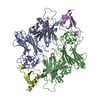
| ||||||||
| 2 | 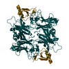
| ||||||||
| Unit cell |
|
-Components
| #1: Protein | Hypothesis / M3 protein Mass: 41826.230 Da / Num. of mol.: 3 Source method: isolated from a genetically manipulated source Source: (gene. exp.) Murid herpesvirus 4 (Murine herpesvirus 68)Genus: Rhadinovirus / Gene: GAMMAHV.M3, M3 / Plasmid: PFB-1 / Cell line (production host): Sf9 / Production host:   Spodoptera frugiperda (fall armyworm) / References: UniProt: O41925 Spodoptera frugiperda (fall armyworm) / References: UniProt: O41925#2: Protein | Mass: 8681.007 Da / Num. of mol.: 3 / Mutation: M87I Source method: isolated from a genetically manipulated source Source: (gene. exp.) Homo sapiens (human) / Gene: CCL2, MCP1, SCYA2 / Plasmid: Paed-4 / Production host:  Escherichia coli (E. coli) / Strain (production host): BL21 PLys S / References: UniProt: P13500 Escherichia coli (E. coli) / Strain (production host): BL21 PLys S / References: UniProt: P13500#3: Water | ChemComp-HOH / | Water Mass: 18.015 Da / Num. of mol.: 562 / Source method: isolated from a natural source / Formula: H2O Mass: 18.015 Da / Num. of mol.: 562 / Source method: isolated from a natural source / Formula: H2O |
|---|
-Experimental details
-Experiment
| Experiment | Method: X-RAY DIFFRACTION / Number of used crystals: 1 |
|---|
- Sample preparation
| Crystal | Density Matthews: 2.28 Å3/Da / Density % sol: 46.11 % |
|---|---|
| Crystal grow | Temperature: 293 K / Method: vapor diffusion, hanging drop / pH: 4.1 Details: 12% PEG 4000, 100 mM sodium acetate, 200 mM magnesium chloride, pH 4.1, VAPOR DIFFUSION, HANGING DROP, temperature 293K |
-Data collection
| Diffraction | Mean temperature: 173 K |
|---|---|
| Diffraction source | Source: SYNCHROTRON / Site: APS  / Beamline: 19-ID / Wavelength: 1 / Beamline: 19-ID / Wavelength: 1 |
| Detector | Type: ADSC QUANTUM 315 / Detector: CCD / Date: Jun 22, 2001 |
| Radiation | Protocol: SINGLE WAVELENGTH / Monochromatic (M) / Laue (L): M / Scattering type: x-ray |
| Radiation wavelength | Wavelength: 1 Å / Relative weight: 1 |
| Reflection | Resolution: 2.5→20 Å / Num. all: 49106 / Num. obs: 45669 / % possible obs: 93.4 % / Observed criterion σ(F): 0 / Redundancy: 6 % / Biso Wilson estimate: 32.7 Å2 / Rsym value: 0.139 / Net I/σ(I): 11.7 |
| Reflection shell | Resolution: 2.5→2.61 Å / Redundancy: 6 % / Mean I/σ(I) obs: 4.1 / Num. unique all: 6652 / Rsym value: 0.412 / % possible all: 87.3 |
- Processing
| Software |
| ||||||||||||||||||||||||||||||||||||
|---|---|---|---|---|---|---|---|---|---|---|---|---|---|---|---|---|---|---|---|---|---|---|---|---|---|---|---|---|---|---|---|---|---|---|---|---|---|
| Refinement | Method to determine structure: MOLECULAR REPLACEMENT Starting model: PDB CODE 1ML0 Resolution: 2.5→19.75 Å / Rfactor Rfree error: 0.006 / Data cutoff high absF: 445022.33 / Data cutoff low absF: 0 / Isotropic thermal model: RESTRAINED / Cross valid method: THROUGHOUT / σ(F): 0
| ||||||||||||||||||||||||||||||||||||
| Solvent computation | Solvent model: FLAT MODEL / Bsol: 44.4868 Å2 / ksol: 0.334325 e/Å3 | ||||||||||||||||||||||||||||||||||||
| Displacement parameters | Biso mean: 37.4 Å2
| ||||||||||||||||||||||||||||||||||||
| Refine analyze |
| ||||||||||||||||||||||||||||||||||||
| Refinement step | Cycle: LAST / Resolution: 2.5→19.75 Å
| ||||||||||||||||||||||||||||||||||||
| Refine LS restraints |
| ||||||||||||||||||||||||||||||||||||
| LS refinement shell | Resolution: 2.5→2.66 Å / Rfactor Rfree error: 0.019 / Total num. of bins used: 6
| ||||||||||||||||||||||||||||||||||||
| Xplor file |
|
