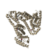
 Movie
Movie Controller


+ Open data

- Basic information
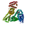
| Entry | Database: PDB / ID: 2i30 | ||||||
|---|---|---|---|---|---|---|---|
| Title | Human serum albumin complexed with myristate and salicylic acid | ||||||
 Components Components | Serum albumin | ||||||
 Keywords Keywords | LIPID BINDING PROTEIN / PLASMA PROTEIN / METAL-BINDING / LIPID-BINDING | ||||||
| Function / homology |  Function and homology information Function and homology informationcellular response to calcium ion starvation / exogenous protein binding / Ciprofloxacin ADME / enterobactin binding / Heme biosynthesis / HDL remodeling / negative regulation of mitochondrial depolarization / Prednisone ADME / Heme degradation / Aspirin ADME ...cellular response to calcium ion starvation / exogenous protein binding / Ciprofloxacin ADME / enterobactin binding / Heme biosynthesis / HDL remodeling / negative regulation of mitochondrial depolarization / Prednisone ADME / Heme degradation / Aspirin ADME / antioxidant activity / toxic substance binding / Scavenging of heme from plasma / Recycling of bile acids and salts / cellular response to starvation / platelet alpha granule lumen / fatty acid binding / Post-translational protein phosphorylation / Cytoprotection by HMOX1 / Regulation of Insulin-like Growth Factor (IGF) transport and uptake by Insulin-like Growth Factor Binding Proteins (IGFBPs) / pyridoxal phosphate binding / Platelet degranulation / protein-folding chaperone binding / blood microparticle / copper ion binding / endoplasmic reticulum lumen / Golgi apparatus / endoplasmic reticulum / protein-containing complex / DNA binding / extracellular space / extracellular exosome / extracellular region / identical protein binding / nucleus / cytoplasmSimilarity search - Function | ||||||
| Biological species |  Homo sapiens (human) Homo sapiens (human) | ||||||
| Method | X-RAY DIFFRACTION / MOLECULAR REPLACEMENT / Resolution: 2.9 Å | ||||||
 Authors Authors | Yang, F. / Bian, C. / Zhu, L. / Zhao, G. / Huang, Z. / Huang, M. | ||||||
 Citation Citation | Journal: J.Struct.Biol. / Year: 2007 Title: Effect of human serum albumin on drug metabolism: Structural evidence of esterase activity of human serum albumin Authors: Yang, F. / Bian, C. / Zhu, L. / Zhao, G. / Huang, Z. / Huang, M. | ||||||
| History |
|
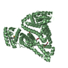
- Structure visualization
| Structure viewer | Molecule: MolmilJmol/JSmol |
|---|
- Downloads & links
-Download
| PDBx/mmCIF format | 2i30.cif.gz | 130.1 KB | Display | PDBx/mmCIF format |
|---|---|---|---|---|
| PDB format | pdb2i30.ent.gz | 100.9 KB | Display | PDB format |
| PDBx/mmJSON format | 2i30.json.gz | Tree view | PDBx/mmJSON format | |
| Others |  Other downloads Other downloads |
-Validation report
| Arichive directory | https://data.pdbj.org/pub/pdb/validation_reports/i3/2i30ftp://data.pdbj.org/pub/pdb/validation_reports/i3/2i30 | HTTPS FTP |
|---|
-Related structure data
| Related structure data |  2i2zC  1bj5S C: citing same article ( S: Starting model for refinement |
|---|---|
| Similar structure data |


-Links
 PDBj
PDBj


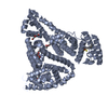
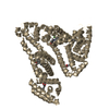
- Assembly
| Deposited unit | 
| ||||||||
|---|---|---|---|---|---|---|---|---|---|
| 1 |
| ||||||||
| Unit cell |
|
-Components
| #1: Protein | Mass: 66571.219 Da / Num. of mol.: 1 / Source method: isolated from a natural source / Source: (natural) Homo sapiens (human) / References: UniProt: P02768 | ||
|---|---|---|---|
| #2: Chemical | Salicylic acid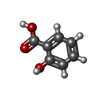  Mass: 138.121 Da / Num. of mol.: 2 / Source method: obtained synthetically / Formula: C7H6O3 Mass: 138.121 Da / Num. of mol.: 2 / Source method: obtained synthetically / Formula: C7H6O3#3: Chemical | ChemComp-MYR / Myristic acid  Mass: 228.371 Da / Num. of mol.: 5 / Source method: obtained synthetically / Formula: C14H28O2 Mass: 228.371 Da / Num. of mol.: 5 / Source method: obtained synthetically / Formula: C14H28O2 |
-Experimental details
-Experiment
| Experiment | Method: X-RAY DIFFRACTION / Number of used crystals: 1 |
|---|
- Sample preparation
| Crystal | Density Matthews: 2.52 Å3/Da / Density % sol: 51.24 % |
|---|---|
| Crystal grow | Temperature: 293 K / Method: evaporation, vapor diffusion, sitting drop / pH: 7.5 Details: 31% PEG 3350, 0.05M potassium phosphate, pH 7.5, EVAPORATION, vapor diffusion, sitting drop, temperature 293K |
-Data collection
| Diffraction | Mean temperature: 293 K |
|---|---|
| Diffraction source | Source: ROTATING ANODE / Type: ENRAF-NONIUS FR591 / Wavelength: 1.5418 / Wavelength: 1.5418 Å |
| Detector | Type: BRUKER SMART 2000 / Detector: CCD / Date: Aug 20, 2004 |
| Radiation | Monochromator: MIRROR / Protocol: SINGLE WAVELENGTH / Monochromatic (M) / Laue (L): M / Scattering type: x-ray |
| Radiation wavelength | Wavelength: 1.5418 Å / Relative weight: 1 |
| Reflection | Resolution: 2.9→45.7 Å / Num. all: 15230 / Num. obs: 13859 / % possible obs: 91 % / Observed criterion σ(F): 2 / Observed criterion σ(I): 6.38 |
| Reflection shell | Resolution: 2.9→3.08 Å / % possible all: 88.7 |
- Processing
| Software |
| ||||||||||||||||||||||||||||
|---|---|---|---|---|---|---|---|---|---|---|---|---|---|---|---|---|---|---|---|---|---|---|---|---|---|---|---|---|---|
| Refinement | Method to determine structure: MOLECULAR REPLACEMENT Starting model: 1BJ5 Resolution: 2.9→45.7 Å / σ(F): 0 / Stereochemistry target values: Engh & Huber
| ||||||||||||||||||||||||||||
| Solvent computation | Bsol: 62.534 Å2 | ||||||||||||||||||||||||||||
| Displacement parameters | Biso mean: 60.788 Å2
| ||||||||||||||||||||||||||||
| Refine analyze |
| ||||||||||||||||||||||||||||
| Refinement step | Cycle: LAST / Resolution: 2.9→45.7 Å
| ||||||||||||||||||||||||||||
| Refine LS restraints |
| ||||||||||||||||||||||||||||
| LS refinement shell | Resolution: 2.9→3.08 Å / Rfactor Rfree error: 0.011
| ||||||||||||||||||||||||||||
| Xplor file |
|
