 Movie
Movie Controller


+ Open data

- Basic information
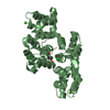
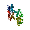
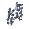
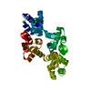
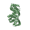
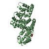
| Entry | Database: PDB / ID: 2hyw | ||||||
|---|---|---|---|---|---|---|---|
| Title | Human Annexin A2 with Calcium bound | ||||||
 Components Components | Annexin A2 | ||||||
 Keywords Keywords | METAL BINDING PROTEIN / calcium-binding protein / membrane-binding protein / helix bundle | ||||||
| Function / homology |  Function and homology information Function and homology informationpositive regulation of low-density lipoprotein particle receptor binding / positive regulation of receptor-mediated endocytosis involved in cholesterol transport / AnxA2-p11 complex / membrane raft assembly / positive regulation of vacuole organization / positive regulation of low-density lipoprotein particle clearance / phospholipase A2 inhibitor activity / positive regulation of vesicle fusion / negative regulation of low-density lipoprotein particle receptor catabolic process / positive regulation of plasma membrane repair ...positive regulation of low-density lipoprotein particle receptor binding / positive regulation of receptor-mediated endocytosis involved in cholesterol transport / AnxA2-p11 complex / membrane raft assembly / positive regulation of vacuole organization / positive regulation of low-density lipoprotein particle clearance / phospholipase A2 inhibitor activity / positive regulation of vesicle fusion / negative regulation of low-density lipoprotein particle receptor catabolic process / positive regulation of plasma membrane repair / positive regulation of plasminogen activation / PCSK9-AnxA2 complex / myelin sheath adaxonal region / cadherin binding involved in cell-cell adhesion / Schmidt-Lanterman incisure / vesicle budding from membrane / cornified envelope / plasma membrane protein complex / calcium-dependent phospholipid binding / negative regulation of receptor internalization / collagen fibril organization / S100 protein binding / Dissolution of Fibrin Clot / virion binding / osteoclast development / positive regulation of low-density lipoprotein receptor activity / epithelial cell apoptotic process / phosphatidylserine binding / positive regulation of receptor recycling / positive regulation of exocytosis / basement membrane / regulation of neurogenesis / Smooth Muscle Contraction / fibrinolysis / phosphatidylinositol-4,5-bisphosphate binding / Gene and protein expression by JAK-STAT signaling after Interleukin-12 stimulation / cytoskeletal protein binding / lipid droplet / cell-matrix adhesion / response to activity / adherens junction / lung development / sarcolemma / mRNA transcription by RNA polymerase II / serine-type endopeptidase inhibitor activity / calcium channel activity / nuclear matrix / RNA polymerase II transcription regulator complex / calcium-dependent protein binding / azurophil granule lumen / melanosome / late endosome membrane / midbody / basolateral plasma membrane / angiogenesis / collagen-containing extracellular matrix / protease binding / vesicle / early endosome / endosome / lysosomal membrane / calcium ion binding / Neutrophil degranulation / cell surface / positive regulation of transcription by RNA polymerase II / extracellular space / RNA binding / extracellular exosome / extracellular region / membrane / identical protein binding / nucleus / plasma membrane / cytosol / cytoplasmSimilarity search - Function | ||||||
| Biological species |  Homo sapiens (human) Homo sapiens (human) | ||||||
| Method | X-RAY DIFFRACTION / MOLECULAR REPLACEMENT / Resolution: 2.1 Å | ||||||
 Authors Authors | Shao, C. / Head, J.F. / Seaton, B.A. | ||||||
 Citation Citation | Journal: J.Biol.Chem. / Year: 2006 Title: Crystallographic Analysis of Calcium-dependent Heparin Binding to Annexin A2. Authors: Shao, C. / Zhang, F. / Kemp, M.M. / Linhardt, R.J. / Waisman, D.M. / Head, J.F. / Seaton, B.A. | ||||||
| History |
|
- Structure visualization
| Structure viewer | Molecule: MolmilJmol/JSmol |
|---|
- Downloads & links
-Download
| PDBx/mmCIF format | 2hyw.cif.gz | 147.5 KB | Display | PDBx/mmCIF format |
|---|---|---|---|---|
| PDB format | pdb2hyw.ent.gz | 113.9 KB | Display | PDB format |
| PDBx/mmJSON format | 2hyw.json.gz | Tree view | PDBx/mmJSON format | |
| Others |  Other downloads Other downloads |
-Validation report
| Arichive directory | https://data.pdbj.org/pub/pdb/validation_reports/hy/2hywftp://data.pdbj.org/pub/pdb/validation_reports/hy/2hyw | HTTPS FTP |
|---|
-Related structure data
| Related structure data |  2hyuC  2hyvC  1xjlS S: Starting model for refinement C: citing same article ( |
|---|---|
| Similar structure data |


-Links
 PDBj
PDBj


- Assembly
| Deposited unit | 
| ||||||||
|---|---|---|---|---|---|---|---|---|---|
| 1 | 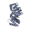
| ||||||||
| 2 | 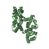
| ||||||||
| Unit cell |
| ||||||||
| Details | The biological unit is a monomer. There are 2 monomers in the asymmetric unit (chains A and B) |
-Components
| #1: Protein | / Annexin II / Lipocortin II / Calpactin I heavy chain / Chromobindin-8 / p36 / Protein I / Placental ...Annexin II / Lipocortin II / Calpactin I heavy chain / Chromobindin-8 / p36 / Protein I / Placental anticoagulant protein IV / PAP-IV Mass: 35352.344 Da / Num. of mol.: 2 Source method: isolated from a genetically manipulated source Source: (gene. exp.) Homo sapiens (human) / Gene: ANXA2 / Plasmid: pAED4.91 / Species (production host): Escherichia coli / Production host:  Escherichia coli BL21(DE3) (bacteria) / Strain (production host): BL21(DE3) / References: UniProt: P07355 Escherichia coli BL21(DE3) (bacteria) / Strain (production host): BL21(DE3) / References: UniProt: P07355#2: Chemical | ChemComp-CA /   Mass: 40.078 Da / Num. of mol.: 14 / Source method: obtained synthetically / Formula: Ca Mass: 40.078 Da / Num. of mol.: 14 / Source method: obtained synthetically / Formula: Ca#3: Water | ChemComp-HOH / | Water Mass: 18.015 Da / Num. of mol.: 438 / Source method: isolated from a natural source / Formula: H2O Mass: 18.015 Da / Num. of mol.: 438 / Source method: isolated from a natural source / Formula: H2O |
|---|
-Experimental details
-Experiment
| Experiment | Method: X-RAY DIFFRACTION / Number of used crystals: 1 |
|---|
- Sample preparation
| Crystal | Density Matthews: 2.86 Å3/Da / Density % sol: 57.04 % |
|---|---|
| Crystal grow | Temperature: 290 K / Method: hanging drop / pH: 8.5 / Details: PEG 8000, pH 8.5, hanging drop, temperature 290K |
-Data collection
| Diffraction |
| ||||||||||||||||||||||||||||||||||||||||||||||||||||||||||||||||||||||||||||||||||||||||||||||||
|---|---|---|---|---|---|---|---|---|---|---|---|---|---|---|---|---|---|---|---|---|---|---|---|---|---|---|---|---|---|---|---|---|---|---|---|---|---|---|---|---|---|---|---|---|---|---|---|---|---|---|---|---|---|---|---|---|---|---|---|---|---|---|---|---|---|---|---|---|---|---|---|---|---|---|---|---|---|---|---|---|---|---|---|---|---|---|---|---|---|---|---|---|---|---|---|---|---|
| Diffraction source | Source: ROTATING ANODE / Type: RIGAKU RU300 / Wavelength: 1.5418 Å | ||||||||||||||||||||||||||||||||||||||||||||||||||||||||||||||||||||||||||||||||||||||||||||||||
| Detector | Type: RIGAKU RAXIS / Detector: IMAGE PLATE / Date: Oct 1, 2004 | ||||||||||||||||||||||||||||||||||||||||||||||||||||||||||||||||||||||||||||||||||||||||||||||||
| Radiation | Protocol: SINGLE WAVELENGTH / Monochromatic (M) / Laue (L): M / Scattering type: x-ray | ||||||||||||||||||||||||||||||||||||||||||||||||||||||||||||||||||||||||||||||||||||||||||||||||
| Radiation wavelength | Wavelength: 1.5418 Å / Relative weight: 1 | ||||||||||||||||||||||||||||||||||||||||||||||||||||||||||||||||||||||||||||||||||||||||||||||||
| Reflection | Resolution: 2.1→40 Å / Num. obs: 45423 / % possible obs: 96.7 % / Redundancy: 3.6 % / Rmerge(I) obs: 0.067 / Χ2: 1.355 / Net I/σ(I): 12.8 | ||||||||||||||||||||||||||||||||||||||||||||||||||||||||||||||||||||||||||||||||||||||||||||||||
| Reflection shell |
|
-Phasing
| Phasing MR | Rfactor: 0.699 / Cor.coef. Fo:Fc: 0.385
|
|---|
- Processing
| Software |
| ||||||||||||||||||||||||||||
|---|---|---|---|---|---|---|---|---|---|---|---|---|---|---|---|---|---|---|---|---|---|---|---|---|---|---|---|---|---|
| Refinement | Method to determine structure: MOLECULAR REPLACEMENT Starting model: PDB entry: 1XJL Resolution: 2.1→40 Å / σ(F): 0
| ||||||||||||||||||||||||||||
| Solvent computation | Bsol: 49.131 Å2 | ||||||||||||||||||||||||||||
| Displacement parameters | Biso mean: 30.068 Å2
| ||||||||||||||||||||||||||||
| Refinement step | Cycle: LAST / Resolution: 2.1→40 Å
| ||||||||||||||||||||||||||||
| Refine LS restraints |
| ||||||||||||||||||||||||||||
| Xplor file |
|
