 Movie
Movie Controller


+ Open data

- Basic information
| Entry | Database: PDB / ID: 2hpl | ||||||
|---|---|---|---|---|---|---|---|
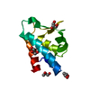
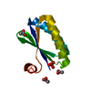
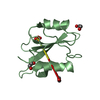
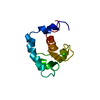
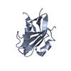
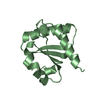
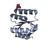
| Title | Crystal structure of the mouse p97/PNGase complex | ||||||
 Components Components |
| ||||||
 Keywords Keywords |  HYDROLASE / PUB domain / Winged helix / p97 HYDROLASE / PUB domain / Winged helix / p97 | ||||||
| Function / homology |  Function and homology informationpeptide-N4-(N-acetyl-beta-glucosaminyl)asparagine amidase / peptide-N4-(N-acetyl-beta-glucosaminyl)asparagine amidase activity / N-glycan trimming in the ER and Calnexin/Calreticulin cycle / glycoprotein catabolic process / protein quality control for misfolded or incompletely synthesized proteins / collagen-containing extracellular matrix / metal ion binding / nucleus / cytosol / cytoplasm Function and homology informationpeptide-N4-(N-acetyl-beta-glucosaminyl)asparagine amidase / peptide-N4-(N-acetyl-beta-glucosaminyl)asparagine amidase activity / N-glycan trimming in the ER and Calnexin/Calreticulin cycle / glycoprotein catabolic process / protein quality control for misfolded or incompletely synthesized proteins / collagen-containing extracellular matrix / metal ion binding / nucleus / cytosol / cytoplasmSimilarity search - Function | ||||||
| Biological species |  Mus musculus (house mouse) Mus musculus (house mouse) | ||||||
| Method | X-RAY DIFFRACTION / SYNCHROTRON / REFMAC 5.2.0005 / Resolution: 1.8 Å | ||||||
 Authors Authors | Zhao, G. / Zhou, X. / Wang, L. / Li, G. / Lennarz, W. / Schindelin, H. | ||||||
 Citation Citation | Journal: Proc.Natl.Acad.Sci.Usa / Year: 2007 Title: Studies on peptide:N-glycanase-p97 interaction suggest that p97 phosphorylation modulates endoplasmic reticulum-associated degradation. Authors: Zhao, G. / Zhou, X. / Wang, L. / Li, G. / Schindelin, H. / Lennarz, W.J. | ||||||
| History |
|
- Structure visualization
| Structure viewer | Molecule: MolmilJmol/JSmol |
|---|
- Downloads & links
-Download
| PDBx/mmCIF format | 2hpl.cif.gz | 36.7 KB | Display | PDBx/mmCIF format |
|---|---|---|---|---|
| PDB format | pdb2hpl.ent.gz | 24.7 KB | Display | PDB format |
| PDBx/mmJSON format | 2hpl.json.gz | Tree view | PDBx/mmJSON format | |
| Others |  Other downloads Other downloads |
-Validation report
| Arichive directory | https://data.pdbj.org/pub/pdb/validation_reports/hp/2hplftp://data.pdbj.org/pub/pdb/validation_reports/hp/2hpl | HTTPS FTP |
|---|
-Related structure data


-Links
 PDBj
PDBj

- Assembly
| Deposited unit | 
| ||||||||
|---|---|---|---|---|---|---|---|---|---|
| 1 |
| ||||||||
| Unit cell |
| ||||||||
| Details | Heterodimer of mouse PNGase PUB and p97 |
-Components
| #1: Protein | Mass: 11198.752 Da / Num. of mol.: 1 Source method: isolated from a genetically manipulated source Source: (gene. exp.) Mus musculus (house mouse) / Gene: Ngly1 / Plasmid: pTYB1 / Production host:  Escherichia coli (E. coli) / Strain (production host): BL21DE3 Codon Plus RIL Escherichia coli (E. coli) / Strain (production host): BL21DE3 Codon Plus RILReferences: UniProt: Q9JI78, peptide-N4-(N-acetyl-beta-glucosaminyl)asparagine amidase |
|---|---|
| #2: Protein/peptide | Mass: 581.574 Da / Num. of mol.: 1 / Source method: obtained synthetically Details: Chemically synthesized. Occurs naturally in mus musculus (mouse) |
| #3: Water | ChemComp-HOH / Water Mass: 18.015 Da / Num. of mol.: 167 / Source method: isolated from a natural source / Formula: H2O Mass: 18.015 Da / Num. of mol.: 167 / Source method: isolated from a natural source / Formula: H2O |
-Experimental details
-Experiment
| Experiment | Method: X-RAY DIFFRACTION / Number of used crystals: 1 |
|---|
- Sample preparation
| Crystal | Density Matthews: 2.31 Å3/Da / Density % sol: 46.75 % |
|---|---|
| Crystal grow | Temperature: 281 K / pH: 8 Details: 18-22% PEG 5000 MME and 14-18% Tacsimate, pH 8.0, VAPOR DIFFUSION, HANGING DROP, temperature 281K |
-Data collection
| Diffraction | Mean temperature: 100 K |
|---|---|
| Diffraction source | Source: SYNCHROTRON / Site: NSLS  / Beamline: X26C / Wavelength: 1.04 Å / Beamline: X26C / Wavelength: 1.04 Å |
| Detector | Type: ADSC QUANTUM 4 / Detector: CCD |
| Radiation | Protocol: SINGLE WAVELENGTH / Monochromatic (M) / Laue (L): M / Scattering type: x-ray |
| Radiation wavelength | Wavelength: 1.04 Å / Relative weight: 1 |
| Reflection | Resolution: 1.8→50 Å / Num. obs: 8764 / % possible obs: 87.7 % / Redundancy: 5.8 % / Rmerge(I) obs: 0.06 / Net I/σ(I): 14.4 |
| Reflection shell | Resolution: 1.8→1.86 Å / Redundancy: 3.8 % / Rmerge(I) obs: 0.314 / % possible all: 46.1 |
- Processing
| Software |
| ||||||||||||||||||||||||||||||||||||||||||||||||||||||||||||||||||||||||||||||||||||||||||||||||||||||||||||||||||||||||||||||||||||||||||||||||||||||||||||||||||||||||||
|---|---|---|---|---|---|---|---|---|---|---|---|---|---|---|---|---|---|---|---|---|---|---|---|---|---|---|---|---|---|---|---|---|---|---|---|---|---|---|---|---|---|---|---|---|---|---|---|---|---|---|---|---|---|---|---|---|---|---|---|---|---|---|---|---|---|---|---|---|---|---|---|---|---|---|---|---|---|---|---|---|---|---|---|---|---|---|---|---|---|---|---|---|---|---|---|---|---|---|---|---|---|---|---|---|---|---|---|---|---|---|---|---|---|---|---|---|---|---|---|---|---|---|---|---|---|---|---|---|---|---|---|---|---|---|---|---|---|---|---|---|---|---|---|---|---|---|---|---|---|---|---|---|---|---|---|---|---|---|---|---|---|---|---|---|---|---|---|---|---|---|---|
| Refinement | Method to determine structure: REFMAC 5.2.0005 / Resolution: 1.8→20 Å / Cor.coef. Fo:Fc: 0.97 / Cor.coef. Fo:Fc free: 0.949 / SU B: 4.459 / SU ML: 0.076 / TLS residual ADP flag: LIKELY RESIDUAL / Cross valid method: THROUGHOUT / σ(F): 0 / ESU R: 0.134 / ESU R Free: 0.123 / Stereochemistry target values: MAXIMUM LIKELIHOOD / Details: HYDROGENS HAVE BEEN ADDED IN THE RIDING POSITIONS
| ||||||||||||||||||||||||||||||||||||||||||||||||||||||||||||||||||||||||||||||||||||||||||||||||||||||||||||||||||||||||||||||||||||||||||||||||||||||||||||||||||||||||||
| Solvent computation | Ion probe radii: 0.8 Å / Shrinkage radii: 0.8 Å / VDW probe radii: 1.2 Å / Solvent model: MASK | ||||||||||||||||||||||||||||||||||||||||||||||||||||||||||||||||||||||||||||||||||||||||||||||||||||||||||||||||||||||||||||||||||||||||||||||||||||||||||||||||||||||||||
| Displacement parameters | Biso mean: 18.76 Å2
| ||||||||||||||||||||||||||||||||||||||||||||||||||||||||||||||||||||||||||||||||||||||||||||||||||||||||||||||||||||||||||||||||||||||||||||||||||||||||||||||||||||||||||
| Refine analyze | Luzzati coordinate error free: 0.242 Å | ||||||||||||||||||||||||||||||||||||||||||||||||||||||||||||||||||||||||||||||||||||||||||||||||||||||||||||||||||||||||||||||||||||||||||||||||||||||||||||||||||||||||||
| Refinement step | Cycle: LAST / Resolution: 1.8→20 Å
| ||||||||||||||||||||||||||||||||||||||||||||||||||||||||||||||||||||||||||||||||||||||||||||||||||||||||||||||||||||||||||||||||||||||||||||||||||||||||||||||||||||||||||
| Refine LS restraints |
| ||||||||||||||||||||||||||||||||||||||||||||||||||||||||||||||||||||||||||||||||||||||||||||||||||||||||||||||||||||||||||||||||||||||||||||||||||||||||||||||||||||||||||
| LS refinement shell | Resolution: 1.8→1.85 Å / Total num. of bins used: 20
| ||||||||||||||||||||||||||||||||||||||||||||||||||||||||||||||||||||||||||||||||||||||||||||||||||||||||||||||||||||||||||||||||||||||||||||||||||||||||||||||||||||||||||
| Refinement TLS params. | Method: refined / Origin x: 40.6055 Å / Origin y: 51.9203 Å / Origin z: 13.9924 Å
| ||||||||||||||||||||||||||||||||||||||||||||||||||||||||||||||||||||||||||||||||||||||||||||||||||||||||||||||||||||||||||||||||||||||||||||||||||||||||||||||||||||||||||
| Refinement TLS group |
|
