 Movie
Movie Controller


+ Open data

- Basic information
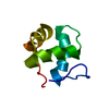
| Entry | Database: PDB / ID: 2hda | ||||||
|---|---|---|---|---|---|---|---|
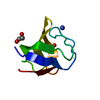
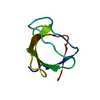
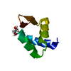
| Title | Yes SH3 domain | ||||||
 Components Components | Proto-oncogene tyrosine-protein kinase Yes | ||||||
 Keywords Keywords |  TRANSFERASE / main beta TRANSFERASE / main beta | ||||||
| Function / homology |  Function and homology information Function and homology informationregulation of glucose transmembrane transport / protein modification process => GO:0036211 / regulation of vascular permeability / CD28 co-stimulation / EPH-Ephrin signaling / microtubule organizing center / epidermal growth factor receptor binding / Regulation of KIT signaling / postsynaptic specialization, intracellular component / CTLA4 inhibitory signaling ...regulation of glucose transmembrane transport / protein modification process => GO:0036211 / regulation of vascular permeability / CD28 co-stimulation / EPH-Ephrin signaling / microtubule organizing center / epidermal growth factor receptor binding / Regulation of KIT signaling / postsynaptic specialization, intracellular component / CTLA4 inhibitory signaling / leukocyte migration / Fc-gamma receptor signaling pathway involved in phagocytosis / EPHA-mediated growth cone collapse / cellular response to platelet-derived growth factor stimulus / RUNX2 regulates osteoblast differentiation / PECAM1 interactions / FCGR activation / EPH-ephrin mediated repulsion of cells / ephrin receptor signaling pathway / cellular response to retinoic acid / peptidyl-tyrosine autophosphorylation / extrinsic component of cytoplasmic side of plasma membrane / Signaling by ERBB2 / cellular response to transforming growth factor beta stimulus / EPHB-mediated forward signaling / T cell costimulation / phosphotyrosine residue binding / FCGR3A-mediated IL10 synthesis / actin filament / Regulation of signaling by CBL / FCGR3A-mediated phagocytosis / cell surface receptor protein tyrosine kinase signaling pathway / non-specific protein-tyrosine kinase / non-membrane spanning protein tyrosine kinase activity / Signaling by SCF-KIT / positive regulation of peptidyl-tyrosine phosphorylation / regulation of cell population proliferation / protein tyrosine kinase activity / transmembrane transporter binding / cell differentiation / signaling receptor binding / focal adhesion / innate immune response / glutamatergic synapse / Golgi apparatus / enzyme binding / positive regulation of transcription by RNA polymerase II / extracellular exosome / ATP binding / plasma membrane / cytosolSimilarity search - Function | ||||||
| Biological species |  Homo sapiens (human) Homo sapiens (human) | ||||||
| Method | X-RAY DIFFRACTION / MOLECULAR REPLACEMENT / Resolution: 1.9 Å | ||||||
 Authors Authors | Camara-Artigas, A. / Luque, I. / Ruiz-Sanz, J. / Mateo, P.L. / Martin-Garcia, J.M. | ||||||
 Citation Citation | Journal: Febs Lett. / Year: 2007 Title: Crystallographic structure of the SH3 domain of the human c-Yes tyrosine kinase: Loop flexibility and amyloid aggregation. Authors: Martin-Garcia, J.M. / Luque, I. / Mateo, P.L. / Ruiz-Sanz, J. / Camara-Artigas, A. | ||||||
| History |
|
- Structure visualization
| Structure viewer | Molecule: MolmilJmol/JSmol |
|---|
- Downloads & links
-Download
| PDBx/mmCIF format | 2hda.cif.gz | 25.4 KB | Display | PDBx/mmCIF format |
|---|---|---|---|---|
| PDB format | pdb2hda.ent.gz | 16.2 KB | Display | PDB format |
| PDBx/mmJSON format | 2hda.json.gz | Tree view | PDBx/mmJSON format | |
| Others |  Other downloads Other downloads |
-Validation report
| Arichive directory | https://data.pdbj.org/pub/pdb/validation_reports/hd/2hdaftp://data.pdbj.org/pub/pdb/validation_reports/hd/2hda | HTTPS FTP |
|---|
-Related structure data
| Related structure data |  1fynS S: Starting model for refinement |
|---|---|
| Similar structure data |


-Links
 PDBj
PDBj


- Assembly
| Deposited unit | 
| ||||||||||||||||||
|---|---|---|---|---|---|---|---|---|---|---|---|---|---|---|---|---|---|---|---|
| 1 |
| ||||||||||||||||||
| 2 | x 6
| ||||||||||||||||||
| 3 | 
| ||||||||||||||||||
| Unit cell |
| ||||||||||||||||||
| Components on special symmetry positions |
|
-Components
| #1: Protein | Mass: 7128.813 Da / Num. of mol.: 1 / Fragment: SH3 domain Source method: isolated from a genetically manipulated source Source: (gene. exp.) Homo sapiens (human) / Gene: YES1, YES / Production host:  Escherichia coli (E. coli) Escherichia coli (E. coli)References: UniProt: P07947, non-specific protein-tyrosine kinase |
|---|---|
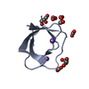
| #2: Chemical | ChemComp-SO4 / Sulfate  Mass: 96.063 Da / Num. of mol.: 1 / Source method: obtained synthetically / Formula: SO4 Mass: 96.063 Da / Num. of mol.: 1 / Source method: obtained synthetically / Formula: SO4 |
| #3: Water | ChemComp-HOH / Water Mass: 18.015 Da / Num. of mol.: 41 / Source method: isolated from a natural source / Formula: H2O Mass: 18.015 Da / Num. of mol.: 41 / Source method: isolated from a natural source / Formula: H2O |
-Experimental details
-Experiment
| Experiment | Method: X-RAY DIFFRACTION / Number of used crystals: 1 |
|---|
- Sample preparation
| Crystal | Density Matthews: 1.99 Å3/Da / Density % sol: 38.19 % |
|---|---|
| Crystal grow | Temperature: 288 K / Method: vapor diffusion, hanging drop / pH: 9 Details: 1.6 M ammonium sulphate, 0.1 M bicine, pH 9.0, VAPOR DIFFUSION, HANGING DROP, temperature 15K, temperature 288K |
-Data collection
| Diffraction | Mean temperature: 100 K |
|---|---|
| Diffraction source | Source: ROTATING ANODE / Type: BRUKER AXS MICROSTAR / Wavelength: 1.54 Å |
| Detector | Type: BRUKER SMART 6500 / Detector: CCD / Date: Jan 30, 2006 / Details: Montel optics |
| Radiation | Monochromator: Bruker Microfocus (Montel Optics) Microstar / Protocol: SINGLE WAVELENGTH / Monochromatic (M) / Laue (L): M / Scattering type: x-ray |
| Radiation wavelength | Wavelength: 1.54 Å / Relative weight: 1 |
| Reflection | Resolution: 1.81→50 Å / Num. obs: 5626 / % possible obs: 99.3 % / Observed criterion σ(F): 0 / Observed criterion σ(I): 0 / Redundancy: 10.08 % / Biso Wilson estimate: 20.3 Å2 / Rmerge(I) obs: 0.0469 / Rsym value: 0.025 / Net I/σ(I): 28.37 |
| Reflection shell | Resolution: 1.81→1.95 Å / Redundancy: 4.19 % / Rmerge(I) obs: 0.238 / Mean I/σ(I) obs: 5.19 / Num. unique all: 1058 / Rsym value: 0.1825 / % possible all: 97.5 |
- Processing
| Software |
| ||||||||||||||||||||||||||||||||||||||||||||||||||||||||||||||||||||||||||||||||||||||||||
|---|---|---|---|---|---|---|---|---|---|---|---|---|---|---|---|---|---|---|---|---|---|---|---|---|---|---|---|---|---|---|---|---|---|---|---|---|---|---|---|---|---|---|---|---|---|---|---|---|---|---|---|---|---|---|---|---|---|---|---|---|---|---|---|---|---|---|---|---|---|---|---|---|---|---|---|---|---|---|---|---|---|---|---|---|---|---|---|---|---|---|---|
| Refinement | Method to determine structure: MOLECULAR REPLACEMENT Starting model: 1FYN Resolution: 1.9→14.86 Å / Cor.coef. Fo:Fc: 0.935 / Cor.coef. Fo:Fc free: 0.891 / SU B: 2.452 / SU ML: 0.076 / Cross valid method: THROUGHOUT / σ(F): 0 / σ(I): 0 / ESU R: 0.186 / ESU R Free: 0.174 / Stereochemistry target values: MAXIMUM LIKELIHOOD / Details: HYDROGENS HAVE BEEN ADDED IN THE RIDING POSITIONS
| ||||||||||||||||||||||||||||||||||||||||||||||||||||||||||||||||||||||||||||||||||||||||||
| Solvent computation | Ion probe radii: 0.8 Å / Shrinkage radii: 0.8 Å / VDW probe radii: 1.4 Å / Solvent model: MASK | ||||||||||||||||||||||||||||||||||||||||||||||||||||||||||||||||||||||||||||||||||||||||||
| Displacement parameters | Biso mean: 20.79 Å2
| ||||||||||||||||||||||||||||||||||||||||||||||||||||||||||||||||||||||||||||||||||||||||||
| Refinement step | Cycle: LAST / Resolution: 1.9→14.86 Å
| ||||||||||||||||||||||||||||||||||||||||||||||||||||||||||||||||||||||||||||||||||||||||||
| Refine LS restraints |
| ||||||||||||||||||||||||||||||||||||||||||||||||||||||||||||||||||||||||||||||||||||||||||
| LS refinement shell | Resolution: 1.9→1.949 Å / Total num. of bins used: 20
|
