 Movie
Movie Controller


+ Open data

- Basic information
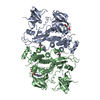
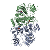
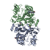
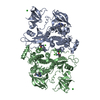
| Entry | Database: PDB / ID: 2hck | ||||||
|---|---|---|---|---|---|---|---|
| Title | SRC FAMILY KINASE HCK-QUERCETIN COMPLEX | ||||||
 Components Components | HEMATOPOETIC CELL KINASE HCK | ||||||
 Keywords Keywords |  TRANSFERASE / PROTEIN TYROSINE KINASE / SIGNAL TRANSDUCTION / SH2 / SH3 TRANSFERASE / PROTEIN TYROSINE KINASE / SIGNAL TRANSDUCTION / SH2 / SH3 | ||||||
| Function / homology |  Function and homology information Function and homology informationleukocyte degranulation / respiratory burst after phagocytosis / innate immune response-activating signaling pathway / leukocyte migration involved in immune response / regulation of podosome assembly / regulation of phagocytosis / FLT3 signaling through SRC family kinases / Nef and signal transduction / regulation of DNA-binding transcription factor activity / positive regulation of actin filament polymerization ...leukocyte degranulation / respiratory burst after phagocytosis / innate immune response-activating signaling pathway / leukocyte migration involved in immune response / regulation of podosome assembly / regulation of phagocytosis / FLT3 signaling through SRC family kinases / Nef and signal transduction / regulation of DNA-binding transcription factor activity / positive regulation of actin filament polymerization / Fc-gamma receptor signaling pathway involved in phagocytosis / mesoderm development / FCGR activation / localization / type II interferon-mediated signaling pathway / Signaling by CSF3 (G-CSF) / lipopolysaccharide-mediated signaling pathway / transport vesicle / negative regulation of inflammatory response to antigenic stimulus / extrinsic component of cytoplasmic side of plasma membrane / phosphotyrosine residue binding / FCGR3A-mediated IL10 synthesis / cell projection / caveola / integrin-mediated signaling pathway / Regulation of signaling by CBL / regulation of actin cytoskeleton organization / FCGR3A-mediated phagocytosis / cell surface receptor protein tyrosine kinase signaling pathway / non-specific protein-tyrosine kinase / non-membrane spanning protein tyrosine kinase activity / Inactivation of CSF3 (G-CSF) signaling / cytoplasmic side of plasma membrane / cytokine-mediated signaling pathway / peptidyl-tyrosine phosphorylation / Signaling by CSF1 (M-CSF) in myeloid cells / regulation of cell shape / regulation of inflammatory response / protein tyrosine kinase activity / protein autophosphorylation / lysosome / cell differentiation / cytoskeleton / cell adhesion / intracellular signal transduction / inflammatory response / protein phosphorylation / signaling receptor binding / intracellular membrane-bounded organelle / focal adhesion / innate immune response / lipid binding / positive regulation of cell population proliferation / negative regulation of apoptotic process / Golgi apparatus / ATP binding / nucleus / plasma membrane / cytosolSimilarity search - Function | ||||||
| Biological species |  Homo sapiens (human) Homo sapiens (human) | ||||||
| Method | X-RAY DIFFRACTION / MIRAS/MAD / Resolution: 3 Å | ||||||
 Authors Authors | Sicheri, F. / Moarefi, I. / Kuriyan, J. | ||||||
 Citation Citation | Journal: Nature / Year: 1997 Title: Crystal structure of the Src family tyrosine kinase Hck. Authors: Sicheri, F. / Moarefi, I. / Kuriyan, J. | ||||||
| History |
|
- Structure visualization
| Structure viewer | Molecule: MolmilJmol/JSmol |
|---|
- Downloads & links
-Download
| PDBx/mmCIF format | 2hck.cif.gz | 212.8 KB | Display | PDBx/mmCIF format |
|---|---|---|---|---|
| PDB format | pdb2hck.ent.gz | 177.8 KB | Display | PDB format |
| PDBx/mmJSON format | 2hck.json.gz | Tree view | PDBx/mmJSON format | |
| Others |  Other downloads Other downloads |
-Validation report
| Arichive directory | https://data.pdbj.org/pub/pdb/validation_reports/hc/2hckftp://data.pdbj.org/pub/pdb/validation_reports/hc/2hck | HTTPS FTP |
|---|
-Related structure data


-Links
 PDBj
PDBj


- Assembly
| Deposited unit | 
| ||||||||
|---|---|---|---|---|---|---|---|---|---|
| 1 |
| ||||||||
| Unit cell |
|
-Components
| #1: Protein | Mass: 50304.348 Da / Num. of mol.: 2 / Fragment: SH3-SH2-KINASE-REGULATORY TAIL Source method: isolated from a genetically manipulated source Source: (gene. exp.) Homo sapiens (human) / Cell line: SF9 / Gene: HUMAN HCK / Cell line (production host): SF9 / Gene (production host): HUMAN HCK / Production host:   Spodoptera frugiperda (fall armyworm) / References: UniProt: P08631, EC: 2.7.1.112 Spodoptera frugiperda (fall armyworm) / References: UniProt: P08631, EC: 2.7.1.112#2: Chemical |   Mass: 40.078 Da / Num. of mol.: 2 / Source method: obtained synthetically / Formula: Ca Mass: 40.078 Da / Num. of mol.: 2 / Source method: obtained synthetically / Formula: Ca#3: Chemical | Quercetin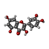  Mass: 302.236 Da / Num. of mol.: 2 / Source method: obtained synthetically / Formula: C15H10O7 Mass: 302.236 Da / Num. of mol.: 2 / Source method: obtained synthetically / Formula: C15H10O7#4: Water | ChemComp-HOH / | Water Mass: 18.015 Da / Num. of mol.: 4 / Source method: isolated from a natural source / Formula: H2O Mass: 18.015 Da / Num. of mol.: 4 / Source method: isolated from a natural source / Formula: H2ONonpolymer details | THE ORIENTATION PLANES OF THE QUERCETIN INHIBITORS IN BOTH HCK MOLECULES OF THE ASYMMETRIC UNIT ARE ...THE ORIENTATIO | |
|---|
-Experimental details
-Experiment
| Experiment | Method: X-RAY DIFFRACTION / Number of used crystals: 1 |
|---|
- Sample preparation
| Crystal | Density Matthews: 3.01 Å3/Da / Density % sol: 59.18 % | ||||||||||||||||||||||||||||||||||||||||||
|---|---|---|---|---|---|---|---|---|---|---|---|---|---|---|---|---|---|---|---|---|---|---|---|---|---|---|---|---|---|---|---|---|---|---|---|---|---|---|---|---|---|---|---|
| Crystal grow | Temperature: 292 K / pH: 6.5 Details: HANGING DROPS (1UL) OF 50MG/ML PROTEIN AND 10MM QUERCETIN WERE MIXED WITH EQUAL VOLUMES OF RESERVOIR BUFFER CONTAINING 150 MM CALCIUM ACETATE, 100MM CACODYLATE (PH 6.5), 7% PEG 8000 AND 16% ...Details: HANGING DROPS (1UL) OF 50MG/ML PROTEIN AND 10MM QUERCETIN WERE MIXED WITH EQUAL VOLUMES OF RESERVOIR BUFFER CONTAINING 150 MM CALCIUM ACETATE, 100MM CACODYLATE (PH 6.5), 7% PEG 8000 AND 16% V/V ETHYLENE GLYCOL. THE MIXED DROPS WERE THEN SEEDED AND STORED AT 19 DEGREES C., temperature 292K | ||||||||||||||||||||||||||||||||||||||||||
| Crystal grow | *PLUS Method: vapor diffusion, hanging dropDetails: drop solution was mixed with an equal volume of reservoir solution | ||||||||||||||||||||||||||||||||||||||||||
| Components of the solutions | *PLUS
|
-Data collection
| Diffraction | Mean temperature: 100 K |
|---|---|
| Diffraction source | Source: ROTATING ANODE / Type: RIGAKU RUH2R / Wavelength: 1.5418 |
| Detector | Type: RIGAKU / Detector: IMAGE PLATE / Date: Nov 1, 1996 / Details: MIRRORS |
| Radiation | Monochromator: NI FILTER / Monochromatic (M) / Laue (L): M / Scattering type: x-ray |
| Radiation wavelength | Wavelength: 1.5418 Å / Relative weight: 1 |
| Reflection | Resolution: 3→18 Å / Num. obs: 385451 / % possible obs: 98.1 % / Observed criterion σ(I): 0 / Redundancy: 15.7 % / Rmerge(I) obs: 0.079 / Rsym value: 0.079 / Net I/σ(I): 17.7 |
| Reflection shell | Resolution: 3→3.11 Å / Redundancy: 6 % / Rmerge(I) obs: 0.145 / Mean I/σ(I) obs: 4.5 / Rsym value: 0.145 / % possible all: 96.4 |
| Reflection | *PLUS Num. obs: 24477 / Num. measured all: 385451 |
| Reflection shell | *PLUS % possible obs: 96.4 % |
- Processing
| Software |
| ||||||||||||||||||||||||||||||||||||||||||||||||||||||||||||
|---|---|---|---|---|---|---|---|---|---|---|---|---|---|---|---|---|---|---|---|---|---|---|---|---|---|---|---|---|---|---|---|---|---|---|---|---|---|---|---|---|---|---|---|---|---|---|---|---|---|---|---|---|---|---|---|---|---|---|---|---|---|
| Refinement | Method to determine structure: MIRAS/MAD / Resolution: 3→18 Å / Data cutoff high absF: 10000000 / Data cutoff low absF: 0.001 / Isotropic thermal model: RESTRAINED / Cross valid method: THROUGHOUT / σ(F): 0
| ||||||||||||||||||||||||||||||||||||||||||||||||||||||||||||
| Displacement parameters | Biso mean: 57.5 Å2
| ||||||||||||||||||||||||||||||||||||||||||||||||||||||||||||
| Refinement step | Cycle: LAST / Resolution: 3→18 Å
| ||||||||||||||||||||||||||||||||||||||||||||||||||||||||||||
| Refine LS restraints |
| ||||||||||||||||||||||||||||||||||||||||||||||||||||||||||||
| Refine LS restraints NCS | NCS model details: RESTRAINTS | ||||||||||||||||||||||||||||||||||||||||||||||||||||||||||||
| LS refinement shell | Resolution: 3→3.08 Å / Total num. of bins used: 13
| ||||||||||||||||||||||||||||||||||||||||||||||||||||||||||||
| Xplor file |
| ||||||||||||||||||||||||||||||||||||||||||||||||||||||||||||
| Software | *PLUS Name: X-PLOR / Version: 3.8 / Classification: refinement | ||||||||||||||||||||||||||||||||||||||||||||||||||||||||||||
| Refinement | *PLUS | ||||||||||||||||||||||||||||||||||||||||||||||||||||||||||||
| Solvent computation | *PLUS | ||||||||||||||||||||||||||||||||||||||||||||||||||||||||||||
| Displacement parameters | *PLUS | ||||||||||||||||||||||||||||||||||||||||||||||||||||||||||||
| LS refinement shell | *PLUS Rfactor obs: 0.355 |
