 Movie
Movie Controller

[English] 日本語
 Yorodumi
- PDB-3vs7: Crystal structure of HCK complexed with a pyrazolo-pyrimidine inh... -
+ Open data

- Basic information
| Entry | Database: PDB / ID: 3vs7 | ||||||
|---|---|---|---|---|---|---|---|
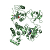
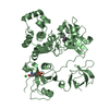
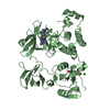
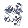
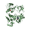
| Title | Crystal structure of HCK complexed with a pyrazolo-pyrimidine inhibitor 1-cyclopentyl-3-(1H-pyrrolo[2,3-b]pyridin-5-yl)-1H-pyrazolo[3,4-d]pyrimidin-4-amine | ||||||
 Components Components | Tyrosine-protein kinase HCK | ||||||
 Keywords Keywords | TRANSFERASE/TRANSFERASE INHIBITOR /  kinase / TRANSFERASE-TRANSFERASE INHIBITOR complex kinase / TRANSFERASE-TRANSFERASE INHIBITOR complex | ||||||
| Function / homology |  Function and homology information Function and homology informationleukocyte degranulation / respiratory burst after phagocytosis / innate immune response-activating signaling pathway / leukocyte migration involved in immune response / regulation of podosome assembly / regulation of phagocytosis / FLT3 signaling through SRC family kinases / Nef and signal transduction / regulation of DNA-binding transcription factor activity / positive regulation of actin filament polymerization ...leukocyte degranulation / respiratory burst after phagocytosis / innate immune response-activating signaling pathway / leukocyte migration involved in immune response / regulation of podosome assembly / regulation of phagocytosis / FLT3 signaling through SRC family kinases / Nef and signal transduction / regulation of DNA-binding transcription factor activity / positive regulation of actin filament polymerization / Fc-gamma receptor signaling pathway involved in phagocytosis / mesoderm development / FCGR activation / localization / type II interferon-mediated signaling pathway / Signaling by CSF3 (G-CSF) / lipopolysaccharide-mediated signaling pathway / transport vesicle / negative regulation of inflammatory response to antigenic stimulus / extrinsic component of cytoplasmic side of plasma membrane / phosphotyrosine residue binding / FCGR3A-mediated IL10 synthesis / cell projection / caveola / integrin-mediated signaling pathway / Regulation of signaling by CBL / regulation of actin cytoskeleton organization / FCGR3A-mediated phagocytosis / cell surface receptor protein tyrosine kinase signaling pathway / non-specific protein-tyrosine kinase / non-membrane spanning protein tyrosine kinase activity / Inactivation of CSF3 (G-CSF) signaling / cytoplasmic side of plasma membrane / cytokine-mediated signaling pathway / peptidyl-tyrosine phosphorylation / Signaling by CSF1 (M-CSF) in myeloid cells / regulation of cell shape / regulation of inflammatory response / protein tyrosine kinase activity / protein autophosphorylation / lysosome / cell differentiation / cytoskeleton / cell adhesion / intracellular signal transduction / inflammatory response / protein phosphorylation / signaling receptor binding / intracellular membrane-bounded organelle / focal adhesion / innate immune response / lipid binding / positive regulation of cell population proliferation / negative regulation of apoptotic process / Golgi apparatus / ATP binding / nucleus / plasma membrane / cytosolSimilarity search - Function | ||||||
| Biological species |  Homo sapiens (human) Homo sapiens (human) | ||||||
| Method | X-RAY DIFFRACTION / SYNCHROTRON / MOLECULAR REPLACEMENT / Resolution: 3.001 Å | ||||||
 Authors Authors | Kuratani, M. / Honda, K. / Niwa, H. / Toyama, M. / Handa, N. / Yokoyama, S. | ||||||
 Citation Citation | Journal: Sci Transl Med / Year: 2013 Title: A Pyrrolo-Pyrimidine Derivative Targets Human Primary AML Stem Cells in Vivo Authors: Saito, Y. / Yuki, H. / Kuratani, M. / Hashizume, Y. / Takagi, S. / Honma, T. / Tanaka, A. / Shirouzu, M. / Mikuni, J. / Handa, N. / Ogahara, I. / Sone, A. / Najima, Y. / Tomabechi, Y. / ...Authors: Saito, Y. / Yuki, H. / Kuratani, M. / Hashizume, Y. / Takagi, S. / Honma, T. / Tanaka, A. / Shirouzu, M. / Mikuni, J. / Handa, N. / Ogahara, I. / Sone, A. / Najima, Y. / Tomabechi, Y. / Wakiyama, M. / Uchida, N. / Tomizawa-Murasawa, M. / Kaneko, A. / Tanaka, S. / Suzuki, N. / Kajita, H. / Aoki, Y. / Ohara, O. / Shultz, L.D. / Fukami, T. / Goto, T. / Taniguchi, S. / Yokoyama, S. / Ishikawa, F. | ||||||
| History |
|
- Structure visualization
| Structure viewer | Molecule: MolmilJmol/JSmol |
|---|
- Downloads & links
-Download
| PDBx/mmCIF format | 3vs7.cif.gz | 188.6 KB | Display | PDBx/mmCIF format |
|---|---|---|---|---|
| PDB format | pdb3vs7.ent.gz | 148.8 KB | Display | PDB format |
| PDBx/mmJSON format | 3vs7.json.gz | Tree view | PDBx/mmJSON format | |
| Others |  Other downloads Other downloads |
-Validation report
| Arichive directory | https://data.pdbj.org/pub/pdb/validation_reports/vs/3vs7ftp://data.pdbj.org/pub/pdb/validation_reports/vs/3vs7 | HTTPS FTP |
|---|
-Related structure data
| Related structure data |  3vrySC  3vrzC  3vs0C  3vs1C  3vs2C  3vs3C  3vs4C  3vs5C  3vs6C S: Starting model for refinement C: citing same article ( |
|---|---|
| Similar structure data |


-Links
 PDBj
PDBj


- Assembly
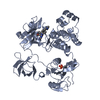
| Deposited unit | 
| ||||||||
|---|---|---|---|---|---|---|---|---|---|
| 1 | 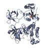
| ||||||||
| 2 | 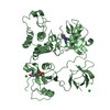
| ||||||||
| Unit cell |
|
-Components
| #1: Protein | Mass: 52000.227 Da / Num. of mol.: 2 / Fragment: UNP residues 81-526 / Mutation: Q528E, Q529E, Q530I Source method: isolated from a genetically manipulated source Source: (gene. exp.) Homo sapiens (human) / Gene: HCK / Production host:   Spodoptera frugiperda (fall armyworm) Spodoptera frugiperda (fall armyworm)References: UniProt: P08631, non-specific protein-tyrosine kinase#2: Chemical |   Mass: 319.364 Da / Num. of mol.: 2 / Source method: obtained synthetically / Formula: C17H17N7 Mass: 319.364 Da / Num. of mol.: 2 / Source method: obtained synthetically / Formula: C17H17N7#3: Chemical |   Mass: 40.078 Da / Num. of mol.: 2 / Source method: obtained synthetically / Formula: Ca Mass: 40.078 Da / Num. of mol.: 2 / Source method: obtained synthetically / Formula: Ca#4: Chemical | Chloride  Mass: 35.453 Da / Num. of mol.: 2 / Source method: obtained synthetically / Formula: Cl Mass: 35.453 Da / Num. of mol.: 2 / Source method: obtained synthetically / Formula: Cl#5: Water | ChemComp-HOH / | Water Mass: 18.015 Da / Num. of mol.: 21 / Source method: isolated from a natural source / Formula: H2O Mass: 18.015 Da / Num. of mol.: 21 / Source method: isolated from a natural source / Formula: H2O |
|---|
-Experimental details
-Experiment
| Experiment | Method: X-RAY DIFFRACTION / Number of used crystals: 1 |
|---|
- Sample preparation
| Crystal | Density Matthews: 3.07 Å3/Da / Density % sol: 59.98 % |
|---|---|
| Crystal grow | Temperature: 277 K / Method: vapor diffusion, sitting drop / pH: 8 Details: 0.1M Tris, 0.1M calcium acetate, 20% glycerol, 21% PEG6000, pH 8, VAPOR DIFFUSION, SITTING DROP, temperature 277K |
-Data collection
| Diffraction | Mean temperature: 100 K |
|---|---|
| Diffraction source | Source: SYNCHROTRON / Site: Photon Factory  / Beamline: BL-1A / Wavelength: 1 Å / Beamline: BL-1A / Wavelength: 1 Å |
| Detector | Type: ADSC QUANTUM 315 / Detector: CCD / Date: Feb 29, 2012 |
| Radiation | Monochromator: Si / Protocol: SINGLE WAVELENGTH / Monochromatic (M) / Laue (L): M / Scattering type: x-ray |
| Radiation wavelength | Wavelength: 1 Å / Relative weight: 1 |
| Reflection | Resolution: 2.95→50 Å / Num. all: 27670 / Num. obs: 27587 / % possible obs: 99.7 % / Observed criterion σ(F): 2 / Observed criterion σ(I): 2 / Biso Wilson estimate: 79.3 Å2 |
| Reflection shell | Resolution: 2.95→3.12 Å / Redundancy: 7.4 % / Mean I/σ(I) obs: 3.8 / % possible all: 99.1 |
- Processing
| Software |
| ||||||||||||||||||||||||||||||||||||||||||||||||||||||||||||||||||||||
|---|---|---|---|---|---|---|---|---|---|---|---|---|---|---|---|---|---|---|---|---|---|---|---|---|---|---|---|---|---|---|---|---|---|---|---|---|---|---|---|---|---|---|---|---|---|---|---|---|---|---|---|---|---|---|---|---|---|---|---|---|---|---|---|---|---|---|---|---|---|---|---|
| Refinement | Method to determine structure: MOLECULAR REPLACEMENT Starting model: 3VRY Resolution: 3.001→42.509 Å / Occupancy max: 1 / Occupancy min: 0.76 / SU ML: 0.34 / σ(F): 1.99 / Phase error: 36.89 / Stereochemistry target values: ML
| ||||||||||||||||||||||||||||||||||||||||||||||||||||||||||||||||||||||
| Solvent computation | Shrinkage radii: 0.86 Å / VDW probe radii: 1.1 Å / Solvent model: FLAT BULK SOLVENT MODEL / Bsol: 68.569 Å2 / ksol: 0.334 e/Å3 | ||||||||||||||||||||||||||||||||||||||||||||||||||||||||||||||||||||||
| Displacement parameters | Biso max: 212.02 Å2 / Biso mean: 108.5404 Å2 / Biso min: 48.65 Å2
| ||||||||||||||||||||||||||||||||||||||||||||||||||||||||||||||||||||||
| Refinement step | Cycle: LAST / Resolution: 3.001→42.509 Å
| ||||||||||||||||||||||||||||||||||||||||||||||||||||||||||||||||||||||
| Refine LS restraints |
| ||||||||||||||||||||||||||||||||||||||||||||||||||||||||||||||||||||||
| LS refinement shell | Refine-ID: X-RAY DIFFRACTION / Total num. of bins used: 9
|
