 Movie
Movie Controller

[English] 日本語
 Yorodumi
- PDB-2hag: Crystal structure of a putative dyp-type peroxidase protein (so_0... -
+ Open data

- Basic information
| Entry | Database: PDB / ID: 2hag | ||||||
|---|---|---|---|---|---|---|---|
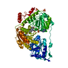
| Title | Crystal structure of a putative dyp-type peroxidase protein (so_0740) from shewanella oneidensis at 2.75 A resolution | ||||||
 Components Components | Melanin biosynthesis protein TyrA, putative | ||||||
 Keywords Keywords |  HYDROLASE / Ferredoxin-like fold / structural genomics / Joint Center for Structural Genomics / JCSG / Protein Structure Initiative / PSI-2 HYDROLASE / Ferredoxin-like fold / structural genomics / Joint Center for Structural Genomics / JCSG / Protein Structure Initiative / PSI-2 | ||||||
| Function / homology |  Function and homology information Function and homology information | ||||||
| Biological species |  Shewanella oneidensis (bacteria) Shewanella oneidensis (bacteria) | ||||||
| Method | X-RAY DIFFRACTION / SYNCHROTRON / MAD / Resolution: 2.75 Å | ||||||
 Authors Authors | Joint Center for Structural Genomics (JCSG) | ||||||
 Citation Citation | Journal: Proteins / Year: 2007 Title: Crystal structures of two novel dye-decolorizing peroxidases reveal a beta-barrel fold with a conserved heme-binding motif. Authors: Zubieta, C. / Krishna, S.S. / Kapoor, M. / Kozbial, P. / McMullan, D. / Axelrod, H.L. / Miller, M.D. / Abdubek, P. / Ambing, E. / Astakhova, T. / Carlton, D. / Chiu, H.J. / Clayton, T. / ...Authors: Zubieta, C. / Krishna, S.S. / Kapoor, M. / Kozbial, P. / McMullan, D. / Axelrod, H.L. / Miller, M.D. / Abdubek, P. / Ambing, E. / Astakhova, T. / Carlton, D. / Chiu, H.J. / Clayton, T. / Deller, M.C. / Duan, L. / Elsliger, M.A. / Feuerhelm, J. / Grzechnik, S.K. / Hale, J. / Hampton, E. / Han, G.W. / Jaroszewski, L. / Jin, K.K. / Klock, H.E. / Knuth, M.W. / Kumar, A. / Marciano, D. / Morse, A.T. / Nigoghossian, E. / Okach, L. / Oommachen, S. / Reyes, R. / Rife, C.L. / Schimmel, P. / van den Bedem, H. / Weekes, D. / White, A. / Xu, Q. / Hodgson, K.O. / Wooley, J. / Deacon, A.M. / Godzik, A. / Lesley, S.A. / Wilson, I.A. | ||||||
| History |
| ||||||
| Remark 999 | SEQUENCE THE CONSTRUCT WAS EXPRESSED WITH A PURIFICATION TAG MGSDKIHHHHHHENLYFQG. THE TAG WAS ...SEQUENCE THE CONSTRUCT WAS EXPRESSED WITH A PURIFICATION TAG MGSDKIHHHHHHENLYFQG. THE TAG WAS REMOVED WITH TEV PROTEASE LEAVING ONLY A GLYCINE (0) FOLLOWED BY THE TARGET SEQUENCE. |
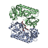
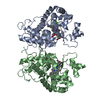
- Structure visualization
| Structure viewer | Molecule: MolmilJmol/JSmol |
|---|
- Downloads & links
-Download
| PDBx/mmCIF format | 2hag.cif.gz | 72.5 KB | Display | PDBx/mmCIF format |
|---|---|---|---|---|
| PDB format | pdb2hag.ent.gz | 57.3 KB | Display | PDB format |
| PDBx/mmJSON format | 2hag.json.gz | Tree view | PDBx/mmJSON format | |
| Others |  Other downloads Other downloads |
-Validation report
| Arichive directory | https://data.pdbj.org/pub/pdb/validation_reports/ha/2hagftp://data.pdbj.org/pub/pdb/validation_reports/ha/2hag | HTTPS FTP |
|---|
-Related structure data


-Links
 PDBj
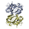
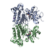
PDBj- Assembly
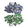
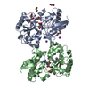
| Deposited unit | 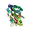
| ||||||||
|---|---|---|---|---|---|---|---|---|---|
| 1 | 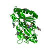
| ||||||||
| Unit cell |
|
-Components
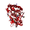
| #1: Protein | Mass: 36469.797 Da / Num. of mol.: 1 Source method: isolated from a genetically manipulated source Source: (gene. exp.) Shewanella oneidensis (bacteria) / Gene: np_716371.1 / Production host: Escherichia coli (E. coli) / References: UniProt: Q8EIU4 |
|---|---|
| #2: Water | ChemComp-HOH / Water Mass: 18.015 Da / Num. of mol.: 48 / Source method: isolated from a natural source / Formula: H2O Mass: 18.015 Da / Num. of mol.: 48 / Source method: isolated from a natural source / Formula: H2O |
-Experimental details
-Experiment
| Experiment | Method: X-RAY DIFFRACTION / Number of used crystals: 1 |
|---|
- Sample preparation
| Crystal | Density Matthews: 3.61 Å3/Da / Density % sol: 65.66 % Description: AFTER THE INITIAL TRACE, A MODEL OF A HOMOLOGOUS PROTEIN (PDB ENTRY 2GVK) WAS USED TO FACILITATE COMPLETION OF THE MODEL. |
|---|---|
| Crystal grow | Temperature: 277 K / Method: vapor diffusion, sitting drop, nanodrop / pH: 7.5 Details: 5.0% iso-Propanol, 20.0% PEG-4000, 0.1M HEPES, pH 7.5, VAPOR DIFFUSION, SITTING DROP, NANODROP, temperature 277K |
-Data collection
| Diffraction | Mean temperature: 100 K | ||||||||||||||||||||||||||||||||||||||||||||||||||||||||||||||||||||||
|---|---|---|---|---|---|---|---|---|---|---|---|---|---|---|---|---|---|---|---|---|---|---|---|---|---|---|---|---|---|---|---|---|---|---|---|---|---|---|---|---|---|---|---|---|---|---|---|---|---|---|---|---|---|---|---|---|---|---|---|---|---|---|---|---|---|---|---|---|---|---|---|
| Diffraction source | Source: SYNCHROTRON / Site: SSRL  / Beamline: BL11-1 / Wavelength: 0.979291, 0.918370, 0.978940 / Beamline: BL11-1 / Wavelength: 0.979291, 0.918370, 0.978940 | ||||||||||||||||||||||||||||||||||||||||||||||||||||||||||||||||||||||
| Detector | Type: ADSC QUANTUM 315 / Detector: CCD / Date: Apr 24, 2006 / Details: Flat mirror (vertical focusing) | ||||||||||||||||||||||||||||||||||||||||||||||||||||||||||||||||||||||
| Radiation | Monochromator: Single crystal Si(111) bent monochromator (horizontal focusing) Protocol: MAD / Monochromatic (M) / Laue (L): M / Scattering type: x-ray | ||||||||||||||||||||||||||||||||||||||||||||||||||||||||||||||||||||||
| Radiation wavelength |
| ||||||||||||||||||||||||||||||||||||||||||||||||||||||||||||||||||||||
| Reflection | Resolution: 2.75→29.54 Å / Num. obs: 13932 / % possible obs: 95.4 % / Biso Wilson estimate: 58.902 Å2 / Rmerge(I) obs: 0.107 / Net I/σ(I): 10.85 | ||||||||||||||||||||||||||||||||||||||||||||||||||||||||||||||||||||||
| Reflection shell | Diffraction-ID: 1
|
-Phasing
| Phasing | Method: MAD |
|---|
- Processing
| Software |
| |||||||||||||||||||||||||||||||||||||||||||||||||||||||||||||||||||||||||||||||||||||||||||||||||||||||||||||||||||||||||||||
|---|---|---|---|---|---|---|---|---|---|---|---|---|---|---|---|---|---|---|---|---|---|---|---|---|---|---|---|---|---|---|---|---|---|---|---|---|---|---|---|---|---|---|---|---|---|---|---|---|---|---|---|---|---|---|---|---|---|---|---|---|---|---|---|---|---|---|---|---|---|---|---|---|---|---|---|---|---|---|---|---|---|---|---|---|---|---|---|---|---|---|---|---|---|---|---|---|---|---|---|---|---|---|---|---|---|---|---|---|---|---|---|---|---|---|---|---|---|---|---|---|---|---|---|---|---|---|
| Refinement | Method to determine structure: MAD / Resolution: 2.75→29.54 Å / Cor.coef. Fo:Fc: 0.948 / Cor.coef. Fo:Fc free: 0.932 / SU B: 22.648 / SU ML: 0.213 / TLS residual ADP flag: LIKELY RESIDUAL / Cross valid method: THROUGHOUT / σ(F): 0 / ESU R: 0.474 / ESU R Free: 0.279 Stereochemistry target values: MAXIMUM LIKELIHOOD WITH PHASES Details: 1. HYDROGENS HAVE BEEN ADDED IN THE RIDING POSITIONS. 2. A MET-INHIBITION PROTOCOL WAS USED FOR SELENOMETHIONINE INCORPORATION DURING PROTEIN EXPRESSION. THE OCCUPANCY OF THE SE ATOMS IN THE ...Details: 1. HYDROGENS HAVE BEEN ADDED IN THE RIDING POSITIONS. 2. A MET-INHIBITION PROTOCOL WAS USED FOR SELENOMETHIONINE INCORPORATION DURING PROTEIN EXPRESSION. THE OCCUPANCY OF THE SE ATOMS IN THE MSE RESIDUES WAS REDUCED TO 0.7 TO ACCOUNT FOR THE REDUCED SCATTERING DUE TO PARTIAL S-MET INCORPORATION. 3. ADDITIONAL ELECTRON DENSITY WAS NOTED ADJACENT TO RESIDUE A20 BUT NOT MODELLED. 4. ATOM RECORD CONTAINS RESIDUAL B FACTORS ONLY.
| |||||||||||||||||||||||||||||||||||||||||||||||||||||||||||||||||||||||||||||||||||||||||||||||||||||||||||||||||||||||||||||
| Solvent computation | Ion probe radii: 0.8 Å / Shrinkage radii: 0.8 Å / VDW probe radii: 1.2 Å / Solvent model: MASK | |||||||||||||||||||||||||||||||||||||||||||||||||||||||||||||||||||||||||||||||||||||||||||||||||||||||||||||||||||||||||||||
| Displacement parameters | Biso mean: 58.485 Å2
| |||||||||||||||||||||||||||||||||||||||||||||||||||||||||||||||||||||||||||||||||||||||||||||||||||||||||||||||||||||||||||||
| Refinement step | Cycle: LAST / Resolution: 2.75→29.54 Å
| |||||||||||||||||||||||||||||||||||||||||||||||||||||||||||||||||||||||||||||||||||||||||||||||||||||||||||||||||||||||||||||
| Refine LS restraints |
| |||||||||||||||||||||||||||||||||||||||||||||||||||||||||||||||||||||||||||||||||||||||||||||||||||||||||||||||||||||||||||||
| LS refinement shell | Resolution: 2.749→2.82 Å / Total num. of bins used: 20
| |||||||||||||||||||||||||||||||||||||||||||||||||||||||||||||||||||||||||||||||||||||||||||||||||||||||||||||||||||||||||||||
| Refinement TLS params. | Method: refined / Origin x: 27.641 Å / Origin y: 27.514 Å / Origin z: 38.523 Å
| |||||||||||||||||||||||||||||||||||||||||||||||||||||||||||||||||||||||||||||||||||||||||||||||||||||||||||||||||||||||||||||
| Refinement TLS group | Selection: ALL |
