 Movie
Movie Controller

[English] 日本語
 Yorodumi
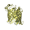
- PDB-2h8i: Crystal Structure of the Bothropstoxin-I complexed with polyethyl... -
+ Open data

- Basic information
| Entry | Database: PDB / ID: 2h8i | ||||||
|---|---|---|---|---|---|---|---|
| Title | Crystal Structure of the Bothropstoxin-I complexed with polyethylene glycol | ||||||
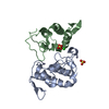
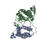
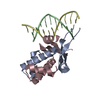
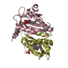
 Components Components | Phospholipase A2 homolog 1 | ||||||
 Keywords Keywords |  TOXIN / Lys49-PLA2s / myotoxicity TOXIN / Lys49-PLA2s / myotoxicity | ||||||
| Function / homology |  Function and homology information Function and homology informationcalcium-dependent phospholipase A2 activity / arachidonic acid secretion / phospholipid metabolic process / lipid catabolic process / negative regulation of T cell proliferation / phospholipid binding / heparin binding / toxin activity / defense response to bacterium / calcium ion binding / extracellular regionSimilarity search - Function | ||||||
| Biological species |  Bothrops jararacussu (jararacussu) Bothrops jararacussu (jararacussu) | ||||||
| Method | X-RAY DIFFRACTION / SYNCHROTRON / MOLECULAR REPLACEMENT / Resolution: 1.9 Å | ||||||
 Authors Authors | Murakami, M.T. / Arni, R.K. | ||||||
 Citation Citation | Journal: Toxicon / Year: 2007 Title: Interfacial surface charge and free accessibility to the PLA2-active site-like region are essential requirements for the activity of Lys49 PLA2 homologues Authors: Murakami, M.T. / Vicotia, M.M. / Abrego, J.R.B. / Lourenzoni, M.R. / Cintra, A.C.O. / Arruda, E.Z. / Tomaz, M.A. / Melo, P.A. / Arni, R.K. | ||||||
| History |
| ||||||
| Remark 999 | sequence The 21th residue is TYR and is a variant present in nature. The 58th and 120th residues ...sequence The 21th residue is TYR and is a variant present in nature. The 58th and 120th residues are ASN and PRO respectively according to Cintra A.C.O. et al.[J. Protein Chem. 12: 57-64(1993)]. |
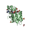
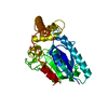
- Structure visualization
| Structure viewer | Molecule: MolmilJmol/JSmol |
|---|
- Downloads & links
-Download
| PDBx/mmCIF format | 2h8i.cif.gz | 58.9 KB | Display | PDBx/mmCIF format |
|---|---|---|---|---|
| PDB format | pdb2h8i.ent.gz | 47.2 KB | Display | PDB format |
| PDBx/mmJSON format | 2h8i.json.gz | Tree view | PDBx/mmJSON format | |
| Others |  Other downloads Other downloads |
-Validation report
| Arichive directory | https://data.pdbj.org/pub/pdb/validation_reports/h8/2h8iftp://data.pdbj.org/pub/pdb/validation_reports/h8/2h8i | HTTPS FTP |
|---|
-Related structure data
| Similar structure data |
|---|


-Links
 PDBj
PDBj
- Assembly
| Deposited unit | 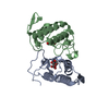
| ||||||||
|---|---|---|---|---|---|---|---|---|---|
| 1 |
| ||||||||
| Unit cell |
| ||||||||
| Details | The biological assembly is the dimer formed in the asymmetric unit. |
-Components
| #1: Protein | Mass: 13752.150 Da / Num. of mol.: 2 / Source method: isolated from a natural source / Details: venom gland / Source: (natural) Bothrops jararacussu (jararacussu) / References: UniProt: Q90249#2: Chemical | Diethylene glycol  Mass: 106.120 Da / Num. of mol.: 2 / Source method: obtained synthetically / Formula: C4H10O3 Mass: 106.120 Da / Num. of mol.: 2 / Source method: obtained synthetically / Formula: C4H10O3#3: Water | ChemComp-HOH / | Water Mass: 18.015 Da / Num. of mol.: 122 / Source method: isolated from a natural source / Formula: H2O Mass: 18.015 Da / Num. of mol.: 122 / Source method: isolated from a natural source / Formula: H2O |
|---|
-Experimental details
-Experiment
| Experiment | Method: X-RAY DIFFRACTION / Number of used crystals: 1 |
|---|
- Sample preparation
| Crystal | Density Matthews: 2.1 Å3/Da / Density % sol: 41.43 % |
|---|---|
| Crystal grow | Temperature: 293 K / Method: vapor diffusion, hanging drop / pH: 7.5 Details: 20% PEG 400, 2M ammonium sulphate, 0.1M HEPES, pH 7.5, VAPOR DIFFUSION, HANGING DROP, temperature 293K |
-Data collection
| Diffraction | Mean temperature: 100 K |
|---|---|
| Diffraction source | Source: SYNCHROTRON / Site: LNLS  / Beamline: D03B-MX1 / Wavelength: 1.438 Å / Beamline: D03B-MX1 / Wavelength: 1.438 Å |
| Detector | Type: MAR CCD 165 mm / Detector: CCD / Date: Jun 20, 2005 |
| Radiation | Protocol: SINGLE WAVELENGTH / Monochromatic (M) / Laue (L): M / Scattering type: x-ray |
| Radiation wavelength | Wavelength: 1.438 Å / Relative weight: 1 |
| Reflection | Resolution: 1.9→40 Å / Num. all: 21766 / Num. obs: 18722 / % possible obs: 99.3 % / Observed criterion σ(F): 2 / Observed criterion σ(I): 3 / Rmerge(I) obs: 0.053 |
| Reflection shell | Resolution: 1.9→1.94 Å / % possible all: 98.8 |
- Processing
| Software |
| ||||||||||||||||||||||||||||||||||||||||||||||||||||||||||||||||||||||||||||||||||||||||||
|---|---|---|---|---|---|---|---|---|---|---|---|---|---|---|---|---|---|---|---|---|---|---|---|---|---|---|---|---|---|---|---|---|---|---|---|---|---|---|---|---|---|---|---|---|---|---|---|---|---|---|---|---|---|---|---|---|---|---|---|---|---|---|---|---|---|---|---|---|---|---|---|---|---|---|---|---|---|---|---|---|---|---|---|---|---|---|---|---|---|---|---|
| Refinement | Method to determine structure: MOLECULAR REPLACEMENT / Resolution: 1.9→10 Å / Cor.coef. Fo:Fc: 0.943 / Cor.coef. Fo:Fc free: 0.927 / SU B: 8.37 / SU ML: 0.128 / TLS residual ADP flag: LIKELY RESIDUAL / Cross valid method: THROUGHOUT / ESU R: 0.201 / ESU R Free: 0.182 / Stereochemistry target values: MAXIMUM LIKELIHOOD
| ||||||||||||||||||||||||||||||||||||||||||||||||||||||||||||||||||||||||||||||||||||||||||
| Solvent computation | Ion probe radii: 0.8 Å / Shrinkage radii: 0.8 Å / VDW probe radii: 1.2 Å / Solvent model: BABINET MODEL WITH MASK | ||||||||||||||||||||||||||||||||||||||||||||||||||||||||||||||||||||||||||||||||||||||||||
| Displacement parameters | Biso mean: 41.481 Å2
| ||||||||||||||||||||||||||||||||||||||||||||||||||||||||||||||||||||||||||||||||||||||||||
| Refinement step | Cycle: LAST / Resolution: 1.9→10 Å
| ||||||||||||||||||||||||||||||||||||||||||||||||||||||||||||||||||||||||||||||||||||||||||
| Refine LS restraints |
| ||||||||||||||||||||||||||||||||||||||||||||||||||||||||||||||||||||||||||||||||||||||||||
| LS refinement shell | Resolution: 1.9→1.948 Å / Total num. of bins used: 20
| ||||||||||||||||||||||||||||||||||||||||||||||||||||||||||||||||||||||||||||||||||||||||||
| Refinement TLS params. | Method: refined / Origin x: 0.7898 Å / Origin y: 23.3794 Å / Origin z: 52.1856 Å
| ||||||||||||||||||||||||||||||||||||||||||||||||||||||||||||||||||||||||||||||||||||||||||
| Refinement TLS group |
|
