 Movie
Movie Controller


+ Open data

- Basic information
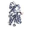
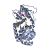
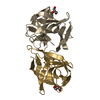
| Entry | Database: PDB / ID: 2h2g | ||||||
|---|---|---|---|---|---|---|---|
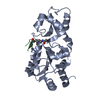
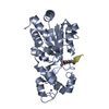
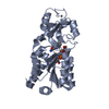
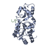
| Title | The Structural Basis of Sirtuin substrate affinity | ||||||
 Components Components |
| ||||||
 Keywords Keywords |  HYDROLASE / H3-K115AC / SIR2 / SIR2TM / SIRTUIN / SIRT1 / HISTONE H3 HYDROLASE / H3-K115AC / SIR2 / SIR2TM / SIRTUIN / SIRT1 / HISTONE H3 | ||||||
| Function / homology |  Function and homology information Function and homology informationsexual sporulation resulting in formation of a cellular spore / global genome nucleotide-excision repair / RNA polymerase I upstream activating factor complex / protein acetyllysine N-acetyltransferase / NAD-dependent histone deacetylase activity / replication fork protection complex / positive regulation of transcription by RNA polymerase I / nucleolar large rRNA transcription by RNA polymerase I / rRNA transcription / NAD+ binding ...sexual sporulation resulting in formation of a cellular spore / global genome nucleotide-excision repair / RNA polymerase I upstream activating factor complex / protein acetyllysine N-acetyltransferase / NAD-dependent histone deacetylase activity / replication fork protection complex / positive regulation of transcription by RNA polymerase I / nucleolar large rRNA transcription by RNA polymerase I / rRNA transcription / NAD+ binding / CENP-A containing nucleosome / structural constituent of chromatin / nucleosome / chromatin organization / transferase activity / protein heterodimerization activity / regulation of DNA-templated transcription / DNA binding / zinc ion binding / nucleus / cytoplasmSimilarity search - Function | ||||||
| Biological species |   Thermotoga maritima (bacteria) Thermotoga maritima (bacteria) | ||||||
| Method | X-RAY DIFFRACTION / SYNCHROTRON / MOLECULAR REPLACEMENT / Resolution: 1.63 Å | ||||||
 Authors Authors | Cosgrove, M.S. / Wolberger, C. | ||||||
 Citation Citation | Journal: Biochemistry / Year: 2006 Title: The structural basis of sirtuin substrate affinity Authors: Cosgrove, M.S. / Bever, K. / Avalos, J.L. / Muhammad, S. / Zhang, X. / Wolberger, C. | ||||||
| History |
|
- Structure visualization
| Structure viewer | Molecule: MolmilJmol/JSmol |
|---|
- Downloads & links
-Download
| PDBx/mmCIF format | 2h2g.cif.gz | 63.9 KB | Display | PDBx/mmCIF format |
|---|---|---|---|---|
| PDB format | pdb2h2g.ent.gz | 49.4 KB | Display | PDB format |
| PDBx/mmJSON format | 2h2g.json.gz | Tree view | PDBx/mmJSON format | |
| Others |  Other downloads Other downloads |
-Validation report
| Arichive directory | https://data.pdbj.org/pub/pdb/validation_reports/h2/2h2gftp://data.pdbj.org/pub/pdb/validation_reports/h2/2h2g | HTTPS FTP |
|---|
-Related structure data


-Links
 PDBj
PDBj


- Assembly
| Deposited unit | 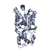
| ||||||||
|---|---|---|---|---|---|---|---|---|---|
| 1 |
| ||||||||
| 2 | 
| ||||||||
| Unit cell |
|
-Components
| #1: Protein | Mass: 27569.793 Da / Num. of mol.: 1 Source method: isolated from a genetically manipulated source Source: (gene. exp.) Thermotoga maritima (bacteria) / Gene: npdA / Production host: Escherichia coli (E. coli)References: UniProt: Q9WYW0, Hydrolases; Acting on carbon-nitrogen bonds, other than peptide bonds; In linear amides | ||
|---|---|---|---|
| #2: Protein/peptide | Mass: 1369.611 Da / Num. of mol.: 1 / Source method: obtained synthetically Details: THE SEQUENCE OF THE PEPTIDE IS NATURALLY FOUND IN SACCHAROMYCES CEREVISIAE (YEAST). References: UniProt: P61830 | ||
| #3: Chemical |   Mass: 65.409 Da / Num. of mol.: 2 / Source method: obtained synthetically / Formula: Zn Mass: 65.409 Da / Num. of mol.: 2 / Source method: obtained synthetically / Formula: Zn#4: Water | ChemComp-HOH / | Water Mass: 18.015 Da / Num. of mol.: 286 / Source method: isolated from a natural source / Formula: H2O Mass: 18.015 Da / Num. of mol.: 286 / Source method: isolated from a natural source / Formula: H2O |
-Experimental details
-Experiment
| Experiment | Method: X-RAY DIFFRACTION / Number of used crystals: 1 |
|---|
- Sample preparation
| Crystal | Density Matthews: 2.56 Å3/Da / Density % sol: 51.98 % |
|---|---|
| Crystal grow | Temperature: 293.15 K / pH: 7.5 Details: 20% PEG, pH 9.6, VAPOR DIFFUSION, HANGING DROP, pH 7.5, temperature 293.15K |
-Data collection
| Diffraction | Mean temperature: 298 K |
|---|---|
| Diffraction source | Source: SYNCHROTRON / Site: APS  / Beamline: 14-BM-D / Wavelength: 1 / Beamline: 14-BM-D / Wavelength: 1 |
| Detector | Type: MARRESEARCH / Detector: IMAGE PLATE / Date: Dec 5, 2004 |
| Radiation | Monochromator: DIAMOND (111) DOUBLE-CRYSTAL MONOCHROMETER / Protocol: SINGLE WAVELENGTH / Monochromatic (M) / Laue (L): M / Scattering type: x-ray |
| Radiation wavelength | Wavelength: 1 Å / Relative weight: 1 |
| Reflection | Resolution: 1.63→50 Å / Num. obs: 37292 / % possible obs: 99.9 % / Observed criterion σ(I): 2 / Redundancy: 6 % / Biso Wilson estimate: 18.8 Å2 / Rmerge(I) obs: 0.069 / Rsym value: 0.045 / Net I/σ(I): 27.1 |
| Reflection shell | Resolution: 1.63→1.69 Å / Redundancy: 6 % / Rmerge(I) obs: 0.514 / Mean I/σ(I) obs: 3.5 / Rsym value: 0.409 / % possible all: 100 |
- Processing
| Software |
| ||||||||||||||||||||||||||||||||||||||||||||||||||||||||||||
|---|---|---|---|---|---|---|---|---|---|---|---|---|---|---|---|---|---|---|---|---|---|---|---|---|---|---|---|---|---|---|---|---|---|---|---|---|---|---|---|---|---|---|---|---|---|---|---|---|---|---|---|---|---|---|---|---|---|---|---|---|---|
| Refinement | Method to determine structure: MOLECULAR REPLACEMENT / Resolution: 1.63→8 Å / Cor.coef. Fo:Fc: 0.951 / Cor.coef. Fo:Fc free: 0.937 / Rfactor Rfree error: 0.005 / SU B: 1.592 / SU ML: 0.057 / Isotropic thermal model: RESTRAINED / Cross valid method: THROUGHOUT / ESU R: 0.093 / ESU R Free: 0.094 / Stereochemistry target values: ENGH & HUBER / Details: HYDROGENS HAVE BEEN ADDED IN THE RIDING POSITIONS
| ||||||||||||||||||||||||||||||||||||||||||||||||||||||||||||
| Solvent computation | Solvent model: FLAT MODEL / Bsol: 85.9797 Å2 / ksol: 0.540206 e/Å3 | ||||||||||||||||||||||||||||||||||||||||||||||||||||||||||||
| Displacement parameters | Biso mean: 20.5 Å2
| ||||||||||||||||||||||||||||||||||||||||||||||||||||||||||||
| Refine analyze |
| ||||||||||||||||||||||||||||||||||||||||||||||||||||||||||||
| Refinement step | Cycle: LAST / Resolution: 1.63→8 Å
| ||||||||||||||||||||||||||||||||||||||||||||||||||||||||||||
| Refine LS restraints |
| ||||||||||||||||||||||||||||||||||||||||||||||||||||||||||||
| LS refinement shell | Resolution: 1.63→1.73 Å / Rfactor Rfree error: 0.017 / Total num. of bins used: 6
|
