 Movie
Movie Controller

[English] 日本語
 Yorodumi
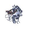
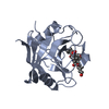
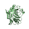
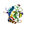
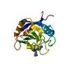
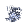
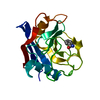
- PDB-2gw2: Crystal structure of the peptidyl-prolyl isomerase domain of huma... -
+ Open data

- Basic information
| Entry | Database: PDB / ID: 2gw2 | ||||||
|---|---|---|---|---|---|---|---|
| Title | Crystal structure of the peptidyl-prolyl isomerase domain of human cyclophilin G | ||||||
 Components Components | Peptidyl-prolyl cis-trans isomerase G | ||||||
 Keywords Keywords |  ISOMERASE / mutation / surface mutagenesis / mutant / ppiase / cis-trans isomerization / peptidyl-prolyl isomerase / protein folding / Structural Genomics / Structural Genomics Consortium / SGC ISOMERASE / mutation / surface mutagenesis / mutant / ppiase / cis-trans isomerization / peptidyl-prolyl isomerase / protein folding / Structural Genomics / Structural Genomics Consortium / SGC | ||||||
| Function / homology |  Function and homology informationcyclosporin A binding / protein peptidyl-prolyl isomerization / mRNA Splicing - Major Pathway / RNA splicing / peptidylprolyl isomerase / peptidyl-prolyl cis-trans isomerase activity / nuclear matrix / SARS-CoV-1 activates/modulates innate immune responses / protein folding / nuclear speck ...cyclosporin A binding / protein peptidyl-prolyl isomerization / mRNA Splicing - Major Pathway / RNA splicing / peptidylprolyl isomerase / peptidyl-prolyl cis-trans isomerase activity / nuclear matrix / SARS-CoV-1 activates/modulates innate immune responses / protein folding / nuclear speck / RNA binding / nucleoplasm / nucleus / cytosol / cytoplasm Function and homology informationcyclosporin A binding / protein peptidyl-prolyl isomerization / mRNA Splicing - Major Pathway / RNA splicing / peptidylprolyl isomerase / peptidyl-prolyl cis-trans isomerase activity / nuclear matrix / SARS-CoV-1 activates/modulates innate immune responses / protein folding / nuclear speck ...cyclosporin A binding / protein peptidyl-prolyl isomerization / mRNA Splicing - Major Pathway / RNA splicing / peptidylprolyl isomerase / peptidyl-prolyl cis-trans isomerase activity / nuclear matrix / SARS-CoV-1 activates/modulates innate immune responses / protein folding / nuclear speck / RNA binding / nucleoplasm / nucleus / cytosol / cytoplasmSimilarity search - Function | ||||||
| Biological species |  Homo sapiens (human) Homo sapiens (human) | ||||||
| Method | X-RAY DIFFRACTION / MOLECULAR REPLACEMENT / Resolution: 1.8 Å | ||||||
 Authors Authors | Bernstein, G. / Tempel, W. / Davis, T. / Newman, E.M. / Finerty Jr., P.J. / Mackenzie, F. / Weigelt, J. / Sundstrom, M. / Arrowsmith, C.H. / Edwards, A.M. ...Bernstein, G. / Tempel, W. / Davis, T. / Newman, E.M. / Finerty Jr., P.J. / Mackenzie, F. / Weigelt, J. / Sundstrom, M. / Arrowsmith, C.H. / Edwards, A.M. / Bochkarev, A. / Dhe-Paganon, S. / Structural Genomics Consortium (SGC) | ||||||
 Citation Citation | Journal: PLoS Biol. / Year: 2010 Title: Structural and biochemical characterization of the human cyclophilin family of peptidyl-prolyl isomerases. Authors: Davis, T.L. / Walker, J.R. / Campagna-Slater, V. / Finerty, P.J. / Paramanathan, R. / Bernstein, G. / MacKenzie, F. / Tempel, W. / Ouyang, H. / Lee, W.H. / Eisenmesser, E.Z. / Dhe-Paganon, S. | ||||||
| History |
| ||||||
| Remark 300 | BIOMOLECULE: THIS ENTRY CONTAINS THE CRYSTALLOGRAPHIC ASYMMETRIC UNIT WHICH CONSISTS OF 1 CHAIN(S). ...BIOMOLECULE: THIS ENTRY CONTAINS THE CRYSTALLOGRAPHIC ASYMMETRIC UNIT WHICH CONSISTS OF 1 CHAIN(S). THE BIOLOGICAL MOLECULE FOR THE PROTEIN IS UNKNOWN. |
- Structure visualization
| Structure viewer | Molecule: MolmilJmol/JSmol |
|---|
- Downloads & links
-Download
| PDBx/mmCIF format | 2gw2.cif.gz | 51.4 KB | Display | PDBx/mmCIF format |
|---|---|---|---|---|
| PDB format | pdb2gw2.ent.gz | 34.8 KB | Display | PDB format |
| PDBx/mmJSON format | 2gw2.json.gz | Tree view | PDBx/mmJSON format | |
| Others |  Other downloads Other downloads |
-Validation report
| Arichive directory | https://data.pdbj.org/pub/pdb/validation_reports/gw/2gw2ftp://data.pdbj.org/pub/pdb/validation_reports/gw/2gw2 | HTTPS FTP |
|---|
-Related structure data
| Related structure data |  1zkcC  2eslC  2he9C 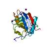 2hq6C 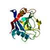 2r99C  1a58S S: Starting model for refinement C: citing same article ( |
|---|---|
| Similar structure data |


-Links
 PDBj
PDBj


- Assembly
| Deposited unit | 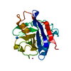
| ||||||||
|---|---|---|---|---|---|---|---|---|---|
| 1 |
| ||||||||
| Unit cell |
| ||||||||
| Details | not known |
-Components
| #1: Protein | Mass: 21842.746 Da / Num. of mol.: 1 / Fragment: residues 1-179 / Mutation: K106A, E107A Source method: isolated from a genetically manipulated source Source: (gene. exp.) Homo sapiens (human) / Gene: PPIG / Plasmid: pET28a / Production host:  Escherichia coli (E. coli) / Strain (production host): BL21(DE3)Codon Plus RIL / References: UniProt: Q13427, peptidylprolyl isomerase Escherichia coli (E. coli) / Strain (production host): BL21(DE3)Codon Plus RIL / References: UniProt: Q13427, peptidylprolyl isomerase | ||
|---|---|---|---|
| #2: Chemical | ChemComp-UNX /   Num. of mol.: 4 / Source method: obtained synthetically Num. of mol.: 4 / Source method: obtained synthetically#3: Water | ChemComp-HOH / | Water Mass: 18.015 Da / Num. of mol.: 103 / Source method: isolated from a natural source / Formula: H2O Mass: 18.015 Da / Num. of mol.: 103 / Source method: isolated from a natural source / Formula: H2O |
-Experimental details
-Experiment
| Experiment | Method: X-RAY DIFFRACTION / Number of used crystals: 1 |
|---|
- Sample preparation
| Crystal | Density Matthews: 1.95 Å3/Da / Density % sol: 36.77 % |
|---|---|
| Crystal grow | Temperature: 291 K / Method: vapor diffusion, sitting drop / pH: 7.5 Details: 2M ammonium sulfate, 0.2M sodium chloride, 0.1M HEPES, pH 7.5, vapor diffusion, sitting drop, temperature 291K |
-Data collection
| Diffraction | Mean temperature: 100 K | ||||||||||||||||||||||||||||||||||||||||||||||||||||||||||||||||||
|---|---|---|---|---|---|---|---|---|---|---|---|---|---|---|---|---|---|---|---|---|---|---|---|---|---|---|---|---|---|---|---|---|---|---|---|---|---|---|---|---|---|---|---|---|---|---|---|---|---|---|---|---|---|---|---|---|---|---|---|---|---|---|---|---|---|---|---|
| Diffraction source | Source: ROTATING ANODE / Type: RIGAKU FR-E / Wavelength: 1.5418 Å | ||||||||||||||||||||||||||||||||||||||||||||||||||||||||||||||||||
| Detector | Type: RIGAKU RAXIS / Detector: IMAGE PLATE / Date: Apr 28, 2006 | ||||||||||||||||||||||||||||||||||||||||||||||||||||||||||||||||||
| Radiation | Protocol: SINGLE WAVELENGTH / Monochromatic (M) / Laue (L): M / Scattering type: x-ray | ||||||||||||||||||||||||||||||||||||||||||||||||||||||||||||||||||
| Radiation wavelength | Wavelength: 1.5418 Å / Relative weight: 1 | ||||||||||||||||||||||||||||||||||||||||||||||||||||||||||||||||||
| Reflection | Resolution: 1.8→30 Å / Num. obs: 14841 / % possible obs: 90 % / Redundancy: 4 % / Rmerge(I) obs: 0.148 / Χ2: 1.66 / Net I/σ(I): 5.8 | ||||||||||||||||||||||||||||||||||||||||||||||||||||||||||||||||||
| Reflection shell |
|
- Processing
| Software |
| ||||||||||||||||||||||||||||||||||||||||||||||||||||||||||||||||||||||||||||||||||||||||||||||||||||||||||||||||||||||||||||||||||||||||||||||||||||||||||||||||||||||||
|---|---|---|---|---|---|---|---|---|---|---|---|---|---|---|---|---|---|---|---|---|---|---|---|---|---|---|---|---|---|---|---|---|---|---|---|---|---|---|---|---|---|---|---|---|---|---|---|---|---|---|---|---|---|---|---|---|---|---|---|---|---|---|---|---|---|---|---|---|---|---|---|---|---|---|---|---|---|---|---|---|---|---|---|---|---|---|---|---|---|---|---|---|---|---|---|---|---|---|---|---|---|---|---|---|---|---|---|---|---|---|---|---|---|---|---|---|---|---|---|---|---|---|---|---|---|---|---|---|---|---|---|---|---|---|---|---|---|---|---|---|---|---|---|---|---|---|---|---|---|---|---|---|---|---|---|---|---|---|---|---|---|---|---|---|---|---|---|---|---|
| Refinement | Method to determine structure: MOLECULAR REPLACEMENT Starting model: pdb entry 1A58 Resolution: 1.8→29.617 Å / Cor.coef. Fo:Fc: 0.932 / Cor.coef. Fo:Fc free: 0.874 / WRfactor Rfree: 0.241 / WRfactor Rwork: 0.189 / SU B: 4.199 / SU ML: 0.129 / ESU R: 0.175 / ESU R Free: 0.172 Details: HYDROGENS HAVE BEEN ADDED IN THE RIDING POSITIONS. ARP/WARP, Molprobity were also used for the refinement.
| ||||||||||||||||||||||||||||||||||||||||||||||||||||||||||||||||||||||||||||||||||||||||||||||||||||||||||||||||||||||||||||||||||||||||||||||||||||||||||||||||||||||||
| Solvent computation | Ion probe radii: 0.8 Å / Shrinkage radii: 0.8 Å / VDW probe radii: 1.4 Å / Solvent model: MASK BULK SOLVENT | ||||||||||||||||||||||||||||||||||||||||||||||||||||||||||||||||||||||||||||||||||||||||||||||||||||||||||||||||||||||||||||||||||||||||||||||||||||||||||||||||||||||||
| Displacement parameters | Biso mean: 14.14 Å2
| ||||||||||||||||||||||||||||||||||||||||||||||||||||||||||||||||||||||||||||||||||||||||||||||||||||||||||||||||||||||||||||||||||||||||||||||||||||||||||||||||||||||||
| Refinement step | Cycle: LAST / Resolution: 1.8→29.617 Å
| ||||||||||||||||||||||||||||||||||||||||||||||||||||||||||||||||||||||||||||||||||||||||||||||||||||||||||||||||||||||||||||||||||||||||||||||||||||||||||||||||||||||||
| Refine LS restraints |
| ||||||||||||||||||||||||||||||||||||||||||||||||||||||||||||||||||||||||||||||||||||||||||||||||||||||||||||||||||||||||||||||||||||||||||||||||||||||||||||||||||||||||
| LS refinement shell | Refine-ID: X-RAY DIFFRACTION / Total num. of bins used: 20
|
