 Movie
Movie Controller

[English] 日本語
 Yorodumi
- PDB-1zkc: Crystal Structure of the cyclophiln_RING domain of human peptidyl... -
+ Open data

- Basic information
| Entry | Database: PDB / ID: 1zkc | ||||||
|---|---|---|---|---|---|---|---|
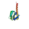
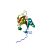
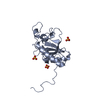
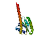
| Title | Crystal Structure of the cyclophiln_RING domain of human peptidylprolyl isomerase (cyclophilin)-like 2 isoform b | ||||||
 Components Components | Peptidyl-prolyl cis-trans isomerase like 2 | ||||||
 Keywords Keywords |  ISOMERASE / CIS-TRANS ISOMERIZATION / peptidylprolyl isomerase / protein-folding / STRUCTURAL GENOMICS CONSORTIUM / SGC ISOMERASE / CIS-TRANS ISOMERIZATION / peptidylprolyl isomerase / protein-folding / STRUCTURAL GENOMICS CONSORTIUM / SGC | ||||||
| Function / homology |  Function and homology information Function and homology informationBasigin interactions / ubiquitin-ubiquitin ligase activity / catalytic step 2 spliceosome / mRNA Splicing - Major Pathway / RNA splicing / protein localization to plasma membrane / RING-type E3 ubiquitin transferase / mRNA processing / Golgi lumen / protein polyubiquitination ...Basigin interactions / ubiquitin-ubiquitin ligase activity / catalytic step 2 spliceosome / mRNA Splicing - Major Pathway / RNA splicing / protein localization to plasma membrane / RING-type E3 ubiquitin transferase / mRNA processing / Golgi lumen / protein polyubiquitination / ubiquitin protein ligase activity / protein folding / nucleoplasm / nucleus / plasma membrane / cytoplasmSimilarity search - Function | ||||||
| Biological species |  Homo sapiens (human) Homo sapiens (human) | ||||||
| Method | X-RAY DIFFRACTION / MOLECULAR REPLACEMENT / Resolution: 1.65 Å | ||||||
 Authors Authors | Walker, J.R. / Davis, T. / Newman, E.M. / Mackenzie, F. / Weigelt, J. / Sundstrom, M. / Arrowsmith, C. / Edwards, A. / Bochkarev, A. / Dhe-Paganon, S. / Structural Genomics Consortium (SGC) | ||||||
 Citation Citation | Journal: PLoS Biol. / Year: 2010 Title: Structural and biochemical characterization of the human cyclophilin family of peptidyl-prolyl isomerases. Authors: Davis, T.L. / Walker, J.R. / Campagna-Slater, V. / Finerty, P.J. / Paramanathan, R. / Bernstein, G. / MacKenzie, F. / Tempel, W. / Ouyang, H. / Lee, W.H. / Eisenmesser, E.Z. / Dhe-Paganon, S. | ||||||
| History |
|
- Structure visualization
| Structure viewer | Molecule: MolmilJmol/JSmol |
|---|
- Downloads & links
-Download
| PDBx/mmCIF format | 1zkc.cif.gz | 180.3 KB | Display | PDBx/mmCIF format |
|---|---|---|---|---|
| PDB format | pdb1zkc.ent.gz | 142.7 KB | Display | PDB format |
| PDBx/mmJSON format | 1zkc.json.gz | Tree view | PDBx/mmJSON format | |
| Others |  Other downloads Other downloads |
-Validation report
| Arichive directory | https://data.pdbj.org/pub/pdb/validation_reports/zk/1zkcftp://data.pdbj.org/pub/pdb/validation_reports/zk/1zkc | HTTPS FTP |
|---|
-Related structure data
| Related structure data |  2eslC  2gw2C  2he9C 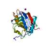 2hq6C  2r99C 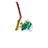 1iipS S: Starting model for refinement C: citing same article ( |
|---|---|
| Similar structure data |


-Links
 PDBj
PDBj


- Assembly
| Deposited unit | 
| ||||||||
|---|---|---|---|---|---|---|---|---|---|
| 1 | 
| ||||||||
| 2 | 
| ||||||||
| Unit cell |
|
-Components
| #1: Protein | Mass: 22168.861 Da / Num. of mol.: 2 / Fragment: sequence databank residues 20-197 Source method: isolated from a genetically manipulated source Source: (gene. exp.) Homo sapiens (human) / Gene: PPIL2 / Plasmid: pET28-LIC / Species (production host): Escherichia coli / Production host:  Escherichia coli BL21(DE3) (bacteria) / Strain (production host): BL21 (DE3) / References: UniProt: Q13356, peptidylprolyl isomerase Escherichia coli BL21(DE3) (bacteria) / Strain (production host): BL21 (DE3) / References: UniProt: Q13356, peptidylprolyl isomerase#2: Chemical | 2-Mercaptoethanol  Mass: 78.133 Da / Num. of mol.: 2 / Source method: obtained synthetically / Formula: C2H6OS Mass: 78.133 Da / Num. of mol.: 2 / Source method: obtained synthetically / Formula: C2H6OS#3: Water | ChemComp-HOH / | Water Mass: 18.015 Da / Num. of mol.: 581 / Source method: isolated from a natural source / Formula: H2O Mass: 18.015 Da / Num. of mol.: 581 / Source method: isolated from a natural source / Formula: H2O |
|---|
-Experimental details
-Experiment
| Experiment | Method: X-RAY DIFFRACTION / Number of used crystals: 1 |
|---|
- Sample preparation
| Crystal | Density Matthews: 2.48 Å3/Da / Density % sol: 49.98 % |
|---|---|
| Crystal grow | Temperature: 298 K / pH: 7.5 Details: 0.8M Potassium Sodium Tartrate tetrahydrate, 0.1M Hepes, pH 7.5, VAPOR DIFFUSION, SITTING DROP, temperature 298K, pH 7.50 |
-Data collection
| Diffraction | Mean temperature: 100 K |
|---|---|
| Diffraction source | Source: ROTATING ANODE / Type: RIGAKU FR-E / Wavelength: 1.5418 |
| Detector | Type: RIGAKU RAXIS IV++ / Detector: IMAGE PLATE / Date: Apr 16, 2005 |
| Radiation | Protocol: SINGLE WAVELENGTH / Monochromatic (M) / Laue (L): M / Scattering type: x-ray |
| Radiation wavelength | Wavelength: 1.5418 Å / Relative weight: 1 |
| Reflection | Resolution: 1.65→30 Å / Num. obs: 57395 / % possible obs: 94.4 % / Observed criterion σ(I): -3 / Redundancy: 5.6 % / Rmerge(I) obs: 0.055 / Net I/σ(I): 31.86 |
| Reflection shell | Resolution: 1.65→1.71 Å / Redundancy: 1.7 % / Rmerge(I) obs: 0.479 / Mean I/σ(I) obs: 1.72 / % possible all: 63.2 |
- Processing
| Software |
| ||||||||||||||||||||||||||||||||||||||||||||||||||||||||||||||||||||||||||||||||||||||||||||||||||||||||||||||||||||||||||||||||||||||||||||||||||||||||||||||||||||||||||
|---|---|---|---|---|---|---|---|---|---|---|---|---|---|---|---|---|---|---|---|---|---|---|---|---|---|---|---|---|---|---|---|---|---|---|---|---|---|---|---|---|---|---|---|---|---|---|---|---|---|---|---|---|---|---|---|---|---|---|---|---|---|---|---|---|---|---|---|---|---|---|---|---|---|---|---|---|---|---|---|---|---|---|---|---|---|---|---|---|---|---|---|---|---|---|---|---|---|---|---|---|---|---|---|---|---|---|---|---|---|---|---|---|---|---|---|---|---|---|---|---|---|---|---|---|---|---|---|---|---|---|---|---|---|---|---|---|---|---|---|---|---|---|---|---|---|---|---|---|---|---|---|---|---|---|---|---|---|---|---|---|---|---|---|---|---|---|---|---|---|---|---|
| Refinement | Method to determine structure: MOLECULAR REPLACEMENT Starting model: PDB ENTRY 1IIP Resolution: 1.65→25.97 Å / Cor.coef. Fo:Fc: 0.967 / Cor.coef. Fo:Fc free: 0.952 / SU B: 3.247 / SU ML: 0.05 / Cross valid method: THROUGHOUT / ESU R: 0.105 / ESU R Free: 0.084 / Stereochemistry target values: MAXIMUM LIKELIHOOD / Details: HYDROGENS HAVE BEEN ADDED IN THE RIDING POSITIONS
| ||||||||||||||||||||||||||||||||||||||||||||||||||||||||||||||||||||||||||||||||||||||||||||||||||||||||||||||||||||||||||||||||||||||||||||||||||||||||||||||||||||||||||
| Solvent computation | Ion probe radii: 0.8 Å / Shrinkage radii: 0.8 Å / VDW probe radii: 1.2 Å / Solvent model: BABINET MODEL WITH MASK | ||||||||||||||||||||||||||||||||||||||||||||||||||||||||||||||||||||||||||||||||||||||||||||||||||||||||||||||||||||||||||||||||||||||||||||||||||||||||||||||||||||||||||
| Displacement parameters | Biso mean: 27.17 Å2
| ||||||||||||||||||||||||||||||||||||||||||||||||||||||||||||||||||||||||||||||||||||||||||||||||||||||||||||||||||||||||||||||||||||||||||||||||||||||||||||||||||||||||||
| Refinement step | Cycle: LAST / Resolution: 1.65→25.97 Å
| ||||||||||||||||||||||||||||||||||||||||||||||||||||||||||||||||||||||||||||||||||||||||||||||||||||||||||||||||||||||||||||||||||||||||||||||||||||||||||||||||||||||||||
| Refine LS restraints |
| ||||||||||||||||||||||||||||||||||||||||||||||||||||||||||||||||||||||||||||||||||||||||||||||||||||||||||||||||||||||||||||||||||||||||||||||||||||||||||||||||||||||||||
| LS refinement shell | Resolution: 1.65→1.693 Å / Total num. of bins used: 20
|
