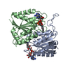
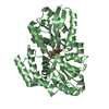
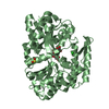
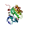
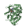
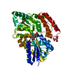
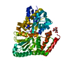
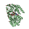
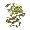
[English] 日本語
Yorodumi
- PDB-2gph: Docking motif interactions in the MAP kinase ERK2 -

+
Open data


ID or keywords:

Loading...

-
Basic information

Entry
Database: PDB / ID: 2gph
TitleDocking motif interactions in the MAP kinase ERK2
Components
  • Mitogen-activated protein kinase 1
  • Tyrosine-protein phosphatase non-receptor type 7
KeywordsTRANSFERASE / ERK2 / docking interaction / D-motif / allostery / CD-site / phosphatase-derived peptide / processing conformation
Function / homology
Function and homology information


phospho-PLA2 pathway / RAF-independent MAPK1/3 activation / MAPK1 (ERK2) activation / Signaling by NODAL / Frs2-mediated activation / ERK/MAPK targets / ERKs are inactivated / Activation of the AP-1 family of transcription factors / RHO GTPases Activate WASPs and WAVEs / IFNG signaling activates MAPKs ...phospho-PLA2 pathway / RAF-independent MAPK1/3 activation / MAPK1 (ERK2) activation / Signaling by NODAL / Frs2-mediated activation / ERK/MAPK targets / ERKs are inactivated / Activation of the AP-1 family of transcription factors / RHO GTPases Activate WASPs and WAVEs / IFNG signaling activates MAPKs / Negative feedback regulation of MAPK pathway / Gastrin-CREB signalling pathway via PKC and MAPK / Estrogen-dependent nuclear events downstream of ESR-membrane signaling / Golgi Cisternae Pericentriolar Stack Reorganization / Regulation of actin dynamics for phagocytic cup formation / Estrogen-stimulated signaling through PRKCZ / Growth hormone receptor signaling / Spry regulation of FGF signaling / SMAD2/SMAD3:SMAD4 heterotrimer regulates transcription / Oxidative Stress Induced Senescence / Senescence-Associated Secretory Phenotype (SASP) / Oncogene Induced Senescence / Signaling by Activin / Downregulation of SMAD2/3:SMAD4 transcriptional activity / Signal attenuation / NCAM signaling for neurite out-growth / Negative regulation of FGFR1 signaling / Negative regulation of FGFR3 signaling / Negative regulation of FGFR4 signaling / Regulation of the apoptosome activity / Signal transduction by L1 / Negative regulation of FGFR2 signaling / RHO GTPases Activate NADPH Oxidases / Negative regulation of MAPK pathway / Interferon gamma signaling / FCERI mediated MAPK activation / PI5P, PP2A and IER3 Regulate PI3K/AKT Signaling / Regulation of HSF1-mediated heat shock response / MAP2K and MAPK activation / diadenosine tetraphosphate biosynthetic process / Recycling pathway of L1 / neural crest cell development / cardiac neural crest cell development involved in heart development / caveolin-mediated endocytosis / cytosine metabolic process / response to epidermal growth factor / mitogen-activated protein kinase kinase kinase binding / regulation of cellular pH / positive regulation of macrophage proliferation / Interleukin-37 signaling / outer ear morphogenesis / Thrombin signalling through proteinase activated receptors (PARs) / RAF/MAP kinase cascade / regulation of Golgi inheritance / ERBB signaling pathway / labyrinthine layer blood vessel development / Neutrophil degranulation / mammary gland epithelial cell proliferation / trachea formation / regulation of early endosome to late endosome transport / : / regulation of stress-activated MAPK cascade / positive regulation of macrophage chemotaxis / lung morphogenesis / ERBB2-ERBB3 signaling pathway / response to exogenous dsRNA / regulation of cytoskeleton organization / face development / androgen receptor signaling pathway / pseudopodium / progesterone receptor signaling pathway / negative regulation of cell differentiation / Bergmann glial cell differentiation / positive regulation of telomere capping / thyroid gland development / decidualization / steroid hormone mediated signaling pathway / non-membrane spanning protein tyrosine phosphatase activity / MAP kinase activity / regulation of ossification / mitogen-activated protein kinase / phosphatase binding / Schwann cell development / stress-activated MAPK cascade / lipopolysaccharide-mediated signaling pathway / positive regulation of cardiac muscle cell proliferation / sensory perception of pain / cellular response to cadmium ion / positive regulation of telomere maintenance via telomerase / ERK1 and ERK2 cascade / cellular response to amino acid starvation / myelination / dendrite cytoplasm / phosphotyrosine residue binding / RNA polymerase II CTD heptapeptide repeat kinase activity / protein dephosphorylation / protein-tyrosine-phosphatase / insulin-like growth factor receptor signaling pathway / thymus development / positive regulation of peptidyl-threonine phosphorylation
Similarity search - Function
Protein-tyrosine phosphatase, KIM-containing / Mitogen-activated protein (MAP) kinase, ERK1/2 / Mitogen-activated protein (MAP) kinase, conserved site / MAP kinase signature. / Protein tyrosine phosphatase, catalytic domain / PTP type protein phosphatase domain profile. / Protein-tyrosine phosphatase / Tyrosine-specific protein phosphatase, PTPase domain / Protein-tyrosine phosphatase, catalytic / Protein tyrosine phosphatase, catalytic domain motif ...Protein-tyrosine phosphatase, KIM-containing / Mitogen-activated protein (MAP) kinase, ERK1/2 / Mitogen-activated protein (MAP) kinase, conserved site / MAP kinase signature. / Protein tyrosine phosphatase, catalytic domain / PTP type protein phosphatase domain profile. / Protein-tyrosine phosphatase / Tyrosine-specific protein phosphatase, PTPase domain / Protein-tyrosine phosphatase, catalytic / Protein tyrosine phosphatase, catalytic domain motif / Tyrosine specific protein phosphatases active site. / Protein-tyrosine phosphatase, active site / Tyrosine-specific protein phosphatases domain / Tyrosine specific protein phosphatases domain profile. / Protein-tyrosine phosphatase-like / Transferase(Phosphotransferase) domain 1 / Transferase(Phosphotransferase); domain 1 / Phosphorylase Kinase; domain 1 / Phosphorylase Kinase; domain 1 / Serine/threonine-protein kinase, active site / Serine/Threonine protein kinases active-site signature. / Protein kinase domain / Serine/Threonine protein kinases, catalytic domain / Protein kinase, ATP binding site / Protein kinases ATP-binding region signature. / Protein kinase domain profile. / Protein kinase domain / Protein kinase-like domain superfamily / 2-Layer Sandwich / Orthogonal Bundle / Mainly Alpha / Alpha Beta
Similarity search - Domain/homology
Tyrosine-protein phosphatase non-receptor type 7 / Mitogen-activated protein kinase 1
Similarity search - Component
Biological speciesRattus norvegicus (Norway rat)
MethodX-RAY DIFFRACTION / SYNCHROTRON / MOLECULAR REPLACEMENT / Resolution: 1.9 Å
AuthorsZhou, T. / Sun, L. / Humphreys, J. / Goldsmith, E.J.
CitationJournal: Structure / Year: 2006
Title: Docking Interactions Induce Exposure of Activation Loop in the MAP Kinase ERK2.
Authors: Zhou, T. / Sun, L. / Humphreys, J. / Goldsmith, E.J.
History
DepositionApr 17, 2006Deposition site: RCSB / Processing site: RCSB
Revision 1.0Jul 4, 2006Provider: repository / Type: Initial release
Revision 1.1May 1, 2008Group: Version format compliance
Revision 1.2Jul 13, 2011Group: Version format compliance
Revision 1.3Oct 18, 2017Group: Refinement description / Category: software
Item: _software.classification / _software.contact_author ..._software.classification / _software.contact_author / _software.contact_author_email / _software.date / _software.language / _software.location / _software.name / _software.type / _software.version
Revision 1.4Oct 20, 2021Group: Database references / Category: database_2 / struct_ref_seq_dif
Item: _database_2.pdbx_DOI / _database_2.pdbx_database_accession / _struct_ref_seq_dif.details
Revision 1.5Aug 30, 2023Group: Data collection / Refinement description
Category: chem_comp_atom / chem_comp_bond / pdbx_initial_refinement_model

-
Structure visualization

Structure viewerMolecule:
MolmilJmol/JSmol

Downloads & links

-
Assembly

Deposited unit
A: Mitogen-activated protein kinase 1
B: Tyrosine-protein phosphatase non-receptor type 7


Theoretical massNumber of molelcules
Total (without water)44,0262
Polymers44,0262
Non-polymers00
Water5,224290
1


  • Idetical with deposited unit
  • defined by author&software
TypeNameSymmetry operationNumber
identity operation1_555x,y,z1
Buried area1540 Å2
ΔGint-10 kcal/mol
Surface area17130 Å2
MethodPISA
Unit cell
Length a, b, c (Å)45.981, 66.342, 116.112
Angle α, β, γ (deg.)90.00, 90.00, 90.00
Int Tables number19
Space group name H-MP212121

-
Components

#1: Protein Mitogen-activated protein kinase 1 / Extracellular signal-regulated kinase 2 / ERK-2 / Mitogen-activated protein kinase 2 / MAP kinase 2 ...Extracellular signal-regulated kinase 2 / ERK-2 / Mitogen-activated protein kinase 2 / MAP kinase 2 / MAPK 2 / p42-MAPK / ERT1


Mass: 42161.473 Da / Num. of mol.: 1 / Mutation: C115T
Source method: isolated from a genetically manipulated source
Source: (gene. exp.) Rattus norvegicus (Norway rat) / Gene: Erk2 / Production host: Escherichia coli (E. coli)
References: UniProt: P63086, mitogen-activated protein kinase
#2: Protein/peptide Tyrosine-protein phosphatase non-receptor type 7 / Protein-tyrosine phosphatase LC-PTP / Hematopoietic protein-tyrosine phosphatase / HEPTP


Mass: 1864.180 Da / Num. of mol.: 1 / Mutation: C52V / Source method: obtained synthetically / Details: This sequence occurs naturally in humans. / References: UniProt: P35236, protein-tyrosine-phosphatase
#3: Water ChemComp-HOH / water / Water


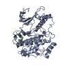
Mass: 18.015 Da / Num. of mol.: 290 / Source method: isolated from a natural source / Formula: H2O

-
Experimental details

-
Experiment

ExperimentMethod: X-RAY DIFFRACTION / Number of used crystals: 1

-
Sample preparation

CrystalDensity Matthews: 2.11 Å3/Da / Density % sol: 41.6 %
Crystal growTemperature: 293 K / Method: vapor diffusion, hanging drop / pH: 7.5
Details: 20% PEG 4000, 10% isopropanol, 0.1M sodium HEPES, pH 7.5, temperature 293K, VAPOR DIFFUSION, HANGING DROP

-
Data collection

DiffractionMean temperature: 90 K
Diffraction sourceSource: SYNCHROTRON / Site: APS / Beamline: 19-BM / Wavelength: 0.97112 Å
DetectorDate: Jan 6, 2005
RadiationProtocol: SINGLE WAVELENGTH / Monochromatic (M) / Laue (L): M / Scattering type: x-ray
Radiation wavelengthWavelength: 0.97112 Å / Relative weight: 1
ReflectionResolution: 1.9→50 Å / Num. obs: 29041

-
Phasing

Phasing dm shell
Resolution (Å)Delta phi finalFOM Reflection
5.56-500.0137.0940.951198
4.41-5.5637.8260.9391192
3.85-4.4130.1890.961148
3.5-3.8536.9080.931171
3.25-3.526.6410.941170
3.06-3.2528.9780.91124
2.9-3.0623.1690.8971127
2.78-2.921.9510.8781119
2.67-2.7815.8750.9271106
2.58-2.6716.3270.8951080
2.5-2.5814.6280.9071124
2.43-2.513.4740.931044
2.36-2.4314.490.8661075
2.31-2.3613.8140.8681042
2.25-2.3114.3050.8971006
2.2-2.2515.0940.8391010
2.16-2.211.20.8781045
2.12-2.1610.4380.891011
2.08-2.1210.0750.889994
2.05-2.0811.9410.841966
2.01-2.0512.5870.842893
1.98-2.019.3890.835829
1.95-1.9810.5290.803796
1.93-1.959.6610.764656
1.9-1.9313.3730.702640

-
Processing

Software
NameVersionClassificationNB
DENZOdata reduction
SCALEPACKdata scaling
CNSrefinement
PDB_EXTRACT1.701data extraction
RefinementMethod to determine structure: MOLECULAR REPLACEMENT
Starting model: PDB Entry: 1ERK

1erk
PDB Unreleased entry


Resolution: 1.9→50 Å / FOM work R set: 0.807 / σ(F): 0
RfactorNum. reflection% reflection
Rfree0.263 1270 4.4 %
Rwork0.216 --
all0.231 --
obs0.231 25566 88.9 %
Solvent computationBsol: 52.789 Å2
Displacement parametersBiso mean: 36.818 Å2
Baniso -1Baniso -2Baniso -3
1-2.357 Å20 Å20 Å2
2--5.586 Å20 Å2
3----7.944 Å2
Refinement stepCycle: LAST / Resolution: 1.9→50 Å
ProteinNucleic acidLigandSolventTotal
Num. atoms2952 0 0 290 3242
Refine LS restraints
Refine-IDTypeDev idealDev ideal target
X-RAY DIFFRACTIONc_mcbond_it1.5461.5
X-RAY DIFFRACTIONc_scbond_it2.3082
X-RAY DIFFRACTIONc_mcangle_it2.4212
X-RAY DIFFRACTIONc_scangle_it3.3732.5
LS refinement shell

Refine-ID: X-RAY DIFFRACTION / Total num. of bins used: 25

Resolution (Å)Rfactor RfreeNum. reflection RfreeRfactor RworkNum. reflection RworkNum. reflection obs
1.9-1.930.338390.414601640
1.93-1.950.432360.365620656
1.95-1.980.394460.328750796
1.98-2.010.306390.323790829
2.01-2.050.345460.302847893
2.05-2.080.29330.296933966
2.08-2.120.261430.258951994
2.12-2.160.289490.2539621011
2.16-2.20.272560.2369891045
2.2-2.250.335460.2939641010
2.25-2.310.258400.2569661006
2.31-2.360.303560.2199861042
2.36-2.430.313490.2210261075
2.43-2.50.207580.1969861044
2.5-2.580.261690.2210551124
2.58-2.670.299510.20310291080
2.67-2.780.223540.19610521106
2.78-2.90.312730.23510461119
2.9-3.060.301460.22610811127
3.06-3.250.299570.21210671124
3.25-3.50.228540.19111161170
3.5-3.850.255490.19911221171
3.85-4.410.203590.15810891148
4.41-5.560.23660.1911261192
5.56-500.010.253560.21211421198
Xplor file
Refine-IDSerial noParam file
X-RAY DIFFRACTION1CNS_TOPPAR:protein_rep.param
X-RAY DIFFRACTION2CNS_TOPPAR:water_rep.param
X-RAY DIFFRACTION3CNS_TOPPAR:ion.param

+
About Yorodumi

-
News

-
Feb 9, 2022. New format data for meta-information of EMDB entries

New format data for meta-information of EMDB entries

  • Version 3 of the EMDB header file is now the official format.
  • The previous official version 1.9 will be removed from the archive.

Related info.:EMDB header

External links:wwPDB to switch to version 3 of the EMDB data model

-
Aug 12, 2020. Covid-19 info

Covid-19 info

URL: https://pdbj.org/emnavi/covid19.php

New page: Covid-19 featured information page in EM Navigator.

Related info.:Covid-19 info / Mar 5, 2020. Novel coronavirus structure data

+
Mar 5, 2020. Novel coronavirus structure data

Novel coronavirus structure data

Related info.:Yorodumi Speices / Aug 12, 2020. Covid-19 info

External links:COVID-19 featured content - PDBj / Molecule of the Month (242):Coronavirus Proteases

+
Jan 31, 2019. EMDB accession codes are about to change! (news from PDBe EMDB page)

EMDB accession codes are about to change! (news from PDBe EMDB page)

  • The allocation of 4 digits for EMDB accession codes will soon come to an end. Whilst these codes will remain in use, new EMDB accession codes will include an additional digit and will expand incrementally as the available range of codes is exhausted. The current 4-digit format prefixed with “EMD-” (i.e. EMD-XXXX) will advance to a 5-digit format (i.e. EMD-XXXXX), and so on. It is currently estimated that the 4-digit codes will be depleted around Spring 2019, at which point the 5-digit format will come into force.
  • The EM Navigator/Yorodumi systems omit the EMD- prefix.

Related info.:Q: What is EMD? / ID/Accession-code notation in Yorodumi/EM Navigator

External links:EMDB Accession Codes are Changing Soon! / Contact to PDBj

+
Jul 12, 2017. Major update of PDB

Major update of PDB

  • wwPDB released updated PDB data conforming to the new PDBx/mmCIF dictionary.
  • This is a major update changing the version number from 4 to 5, and with Remediation, in which all the entries are updated.
  • In this update, many items about electron microscopy experimental information are reorganized (e.g. em_software).
  • Now, EM Navigator and Yorodumi are based on the updated data.

External links:wwPDB Remediation / Enriched Model Files Conforming to OneDep Data Standards Now Available in the PDB FTP Archive

-
Yorodumi

Thousand views of thousand structures

  • Yorodumi is a browser for structure data from EMDB, PDB, SASBDB, etc.
  • This page is also the successor to EM Navigator detail page, and also detail information page/front-end page for Omokage search.
  • The word "yorodu" (or yorozu) is an old Japanese word meaning "ten thousand". "mi" (miru) is to see.

Related info.:EMDB / PDB / SASBDB / Comparison of 3 databanks / Yorodumi Search / Aug 31, 2016. New EM Navigator & Yorodumi / Yorodumi Papers / Jmol/JSmol / Function and homology information / Changes in new EM Navigator and Yorodumi

Read more