 Movie
Movie Controller

[English] 日本語
 Yorodumi
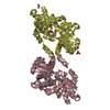
- PDB-2gfp: Structure of the Multidrug Transporter EmrD from Escherichia coli -
+ Open data

- Basic information
| Entry | Database: PDB / ID: 2gfp | ||||||
|---|---|---|---|---|---|---|---|
| Title | Structure of the Multidrug Transporter EmrD from Escherichia coli | ||||||
 Components Components | Multidrug resistance protein D Multiple drug resistance Multiple drug resistance | ||||||
 Keywords Keywords | MEMBRANE PROTEIN / Multidrug Transporter | ||||||
| Function / homology |  Function and homology information Function and homology informationxenobiotic transmembrane transport / xenobiotic detoxification by transmembrane export across the plasma membrane / xenobiotic transmembrane transporter activity / plasma membrane => GO:0005886 / plasma membraneSimilarity search - Function | ||||||
| Biological species |  Escherichia coli (E. coli) Escherichia coli (E. coli) | ||||||
| Method | X-RAY DIFFRACTION / SYNCHROTRON / MAD / Resolution: 3.5 Å | ||||||
 Authors Authors | Yin, Y. / He, X. / Szewczyk, P. / Nguyen, T. / Chang, G. | ||||||
 Citation Citation | Journal: Science / Year: 2006 Title: Structure of the multidrug transporter EmrD from Escherichia coli Authors: Yin, Y. / He, X. / Szewczyk, P. / Nguyen, T. / Chang, G. | ||||||
| History |
|
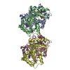
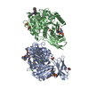
- Structure visualization
| Structure viewer | Molecule: MolmilJmol/JSmol |
|---|
- Downloads & links
-Download
| PDBx/mmCIF format | 2gfp.cif.gz | 127.7 KB | Display | PDBx/mmCIF format |
|---|---|---|---|---|
| PDB format | pdb2gfp.ent.gz | 97.9 KB | Display | PDB format |
| PDBx/mmJSON format | 2gfp.json.gz | Tree view | PDBx/mmJSON format | |
| Others |  Other downloads Other downloads |
-Validation report
| Arichive directory | https://data.pdbj.org/pub/pdb/validation_reports/gf/2gfpftp://data.pdbj.org/pub/pdb/validation_reports/gf/2gfp | HTTPS FTP |
|---|
-Related structure data
| Similar structure data |
|---|


-Links
 PDBj
PDBj


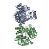
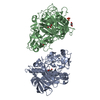
- Assembly
| Deposited unit | 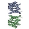
| ||||||||
|---|---|---|---|---|---|---|---|---|---|
| 1 |
| ||||||||
| Unit cell |
|
-Components
| #1: Protein | Multiple drug resistance / EmrD Mass: 40013.898 Da / Num. of mol.: 2 Source method: isolated from a genetically manipulated source Source: (gene. exp.) Escherichia coli (E. coli) / Gene: emrD / Production host: Escherichia coli (E. coli) / References: UniProt: P31442 |
|---|
-Experimental details
-Experiment
| Experiment | Method: X-RAY DIFFRACTION / Number of used crystals: 1 |
|---|
- Sample preparation
| Crystal | Density Matthews: 5.5 Å3/Da / Density % sol: 74.61 % |
|---|---|
| Crystal grow | Method: vapor diffusion, sitting drop / pH: 4.4 Details: 20-21% PEG 400, 50 mM sodium citrate, pH 4.4 and 4.8, and 50 mM KCl, VAPOR DIFFUSION, SITTING DROP |
-Data collection
| Diffraction | Mean temperature: 100 K | |||||||||
|---|---|---|---|---|---|---|---|---|---|---|
| Diffraction source | Source: SYNCHROTRON / Site: ALS  / Beamline: 8.2.1 / Wavelength: 1.03840,1.04040 / Beamline: 8.2.1 / Wavelength: 1.03840,1.04040 | |||||||||
| Detector | Type: ADSC QUANTUM 210 / Detector: CCD | |||||||||
| Radiation | Monochromator: graphite / Protocol: MAD / Monochromatic (M) / Laue (L): M / Scattering type: x-ray | |||||||||
| Radiation wavelength |
| |||||||||
| Reflection | Resolution: 3.5→50 Å / Num. all: 16781 / Num. obs: 13425 / % possible obs: 90 % / Observed criterion σ(I): 1 | |||||||||
| Reflection shell | Resolution: 3.5→3.7 Å / % possible all: 90 |
- Processing
| Software |
| |||||||||||||||||||||||||
|---|---|---|---|---|---|---|---|---|---|---|---|---|---|---|---|---|---|---|---|---|---|---|---|---|---|---|
| Refinement | Method to determine structure: MAD / Resolution: 3.5→50 Å / σ(F): 2 / Stereochemistry target values: Engh & Huber
| |||||||||||||||||||||||||
| Refinement step | Cycle: LAST / Resolution: 3.5→50 Å
| |||||||||||||||||||||||||
| Refine LS restraints |
|
