 Movie
Movie Controller


+ Open data

- Basic information
| Entry | Database: PDB / ID: 2gez | ||||||
|---|---|---|---|---|---|---|---|
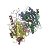
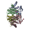
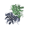
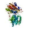
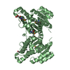
| Title | Crystal structure of potassium-independent plant asparaginase | ||||||
 Components Components |
| ||||||
 Keywords Keywords |  HYDROLASE / isoaspartyl aminopeptidase / L-asparaginase / Ntn-hydrolase / autoproteolysis / taspase / sodium binding HYDROLASE / isoaspartyl aminopeptidase / L-asparaginase / Ntn-hydrolase / autoproteolysis / taspase / sodium binding | ||||||
| Function / homology |  Function and homology information Function and homology information | ||||||
| Biological species |   Lupinus luteus (yellow lupine) Lupinus luteus (yellow lupine) | ||||||
| Method | X-RAY DIFFRACTION / SYNCHROTRON / MOLECULAR REPLACEMENT / Resolution: 2.6 Å | ||||||
 Authors Authors | Michalska, K. / Bujacz, G. / Jaskolski, M. | ||||||
 Citation Citation | Journal: J.Mol.Biol. / Year: 2006 Title: Crystal structure of plant asparaginase. Authors: Michalska, K. / Bujacz, G. / Jaskolski, M. #1: Journal: Eur.J.Biochem. / Year: 2004Title: Expression, purification and catalytic activity of Lupinus luteus asparagine beta-amidohydrolase and its Escherichia coli homolog. Authors: Borek, D. / Michalska, K. / Brzezinski, K. / Kisiel, A. / Podkowinski, J. / Bonthron, T.D. / Krowarsch, D. / Otlewski, J. / Jaskolski, M. #2: Journal: J.Biol.Chem. / Year: 2005Title: Crystal structure of isoaspartyl aminopeptidase in complex with L-aspartate. Authors: Michalska, K. / Brzezinski, K. / Jaskolski, M. #3: Journal: Acta Crystallogr.,Sect.D / Year: 2000Title: Crystallization and preliminary crystallographic studies of a new L-asparaginase encoded by the Escherichia coli genome. Authors: Borek, D. / Jaskolski, M. #4: Journal: Acta Crystallogr.,Sect.D / Year: 2004Title: Structure of the isoaspartyl peptidase with L-asparaginase activity from Escherichia coli. Authors: Prahl, A. / Pazgier, M. / Hejazi, M. / Lockau, W. / Lubkowski, J. #5: Journal: Nature / Year: 1995Title: A protein catalytic framework with an N-terminal nucleophile is capable of self-activation. Authors: Brannigan, J.A. / Dodson, G. / Duggleby, H.J. / Moody, P.C. / Smith, J.L. / Tomchick, D.R. / Murzin, A.G. #6: Journal: Nat.Struct.Biol. / Year: 1995Title: Three-dimensional structure of human lysosomal aspartylglucosaminidase. Authors: Oinonen, C. / Tikkanen, R. / Rouvinen, J. / Peltonen, L. #7: Journal: J.Biol.Chem. / Year: 1998Title: Crystal structures of Flavobacterium glycosylasparaginase. An N-terminal nucleophile hydrolase activated by intramolecular proteolysis. Authors: Guo, H.C. / Xu, Q. / Buckley, D. / Guan, C. #8: Journal: Structure / Year: 2005Title: Crystal structure of human Taspase1, a crucial protease regulating the function of MLL. Authors: Khan, J.A. / Dunn, B.M. / Tong, L. | ||||||
| History |
|
- Structure visualization
| Structure viewer | Molecule: MolmilJmol/JSmol |
|---|
- Downloads & links
-Download
| PDBx/mmCIF format | 2gez.cif.gz | 225.9 KB | Display | PDBx/mmCIF format |
|---|---|---|---|---|
| PDB format | pdb2gez.ent.gz | 179.9 KB | Display | PDB format |
| PDBx/mmJSON format | 2gez.json.gz | Tree view | PDBx/mmJSON format | |
| Others |  Other downloads Other downloads |
-Validation report
| Arichive directory | https://data.pdbj.org/pub/pdb/validation_reports/ge/2gezftp://data.pdbj.org/pub/pdb/validation_reports/ge/2gez | HTTPS FTP |
|---|
-Related structure data
| Related structure data |  1k2xS S: Starting model for refinement |
|---|---|
| Similar structure data |


-Links
 PDBj
PDBj


- Assembly
| Deposited unit | 
| ||||||||
|---|---|---|---|---|---|---|---|---|---|
| 1 | 
| ||||||||
| 2 | 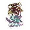
| ||||||||
| Unit cell |
| ||||||||
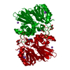
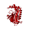
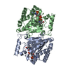
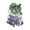
| Details | The asymmetric unit contains two biological assemblies. Each of them is an (alpha/beta)2 heterotetramer or a dimer of (alpha/beta) heterodimers. Subunits alpha (chains A, C, E, G) and beta (chains B, D, F, H) are, respectively, the N- and C-terminal products of autoproteolytic cleavage of a precursor. |
-Components
| #1: Protein | Mass: 21165.877 Da / Num. of mol.: 4 / Fragment: N-terminal subunit (residues 1-192) Source method: isolated from a genetically manipulated source Source: (gene. exp.) Lupinus luteus (yellow lupine) / Strain: cv. Ventus / Plasmid: pET15b / Production host:  Escherichia coli (E. coli) / Strain (production host): BL21 Codon Plus RIP / References: UniProt: Q9ZSD6, asparaginase Escherichia coli (E. coli) / Strain (production host): BL21 Codon Plus RIP / References: UniProt: Q9ZSD6, asparaginase#2: Protein | Mass: 13617.478 Da / Num. of mol.: 4 / Fragment: C-terminal subunit (residues 193-325) Source method: isolated from a genetically manipulated source Details: Subunits alpha (chains A, C, E, G) and beta (chains B, D, F, H) are, respectively, the N- and C-terminal products of autoproteolytic cleavage of a precursor Source: (gene. exp.) Lupinus luteus (yellow lupine) / Strain: cv. Ventus / Plasmid: pET15b / Production host: Escherichia coli (E. coli) / Strain (production host): BL21 Codon Plus RIP / References: UniProt: Q9ZSD6, asparaginase#3: Chemical | ChemComp-NA /   Mass: 22.990 Da / Num. of mol.: 4 / Source method: obtained synthetically / Formula: Na Mass: 22.990 Da / Num. of mol.: 4 / Source method: obtained synthetically / Formula: Na#4: Chemical | ChemComp-CL / Chloride  Mass: 35.453 Da / Num. of mol.: 6 / Source method: obtained synthetically / Formula: Cl Mass: 35.453 Da / Num. of mol.: 6 / Source method: obtained synthetically / Formula: Cl#5: Water | ChemComp-HOH / | Water Mass: 18.015 Da / Num. of mol.: 112 / Source method: isolated from a natural source / Formula: H2O Mass: 18.015 Da / Num. of mol.: 112 / Source method: isolated from a natural source / Formula: H2O |
|---|
-Experimental details
-Experiment
| Experiment | Method: X-RAY DIFFRACTION / Number of used crystals: 1 |
|---|
- Sample preparation
| Crystal | Density Matthews: 2.11 Å3/Da / Density % sol: 41.77 % |
|---|---|
| Crystal grow | Temperature: 292 K / Method: vapor diffusion, hanging drop / pH: 6.5 Details: 20% PEG 4000, 100 mM HEPES, 200 mM MgCl2, pH 6.5, VAPOR DIFFUSION, HANGING DROP, temperature 292.0K |
-Data collection
| Diffraction | Mean temperature: 100 K |
|---|---|
| Diffraction source | Source: SYNCHROTRON / Site: EMBL/DESY, HAMBURG  / Beamline: X11 / Wavelength: 0.81 Å / Beamline: X11 / Wavelength: 0.81 Å |
| Detector | Type: MARRESEARCH / Detector: CCD / Date: Mar 5, 2004 |
| Radiation | Monochromator: Triangular monochromator / Protocol: SINGLE WAVELENGTH / Monochromatic (M) / Laue (L): M / Scattering type: x-ray |
| Radiation wavelength | Wavelength: 0.81 Å / Relative weight: 1 |
| Reflection | Resolution: 2.6→25 Å / Num. all: 34621 / Num. obs: 34621 / % possible obs: 99 % / Observed criterion σ(F): 0 / Observed criterion σ(I): -3 / Redundancy: 2.8 % / Biso Wilson estimate: 40.9 Å2 / Rmerge(I) obs: 0.073 / Net I/σ(I): 14.3 |
| Reflection shell | Resolution: 2.6→2.69 Å / Redundancy: 2.5 % / Rmerge(I) obs: 0.152 / Mean I/σ(I) obs: 6.1 / Num. unique all: 3371 / % possible all: 96.9 |
- Processing
| Software |
| |||||||||||||||||||||||||||||||||||||||||||||||||||||||||||||||||||||||||||||||||||||||||||||||||||||||||||||||||||||||||||||||||||||||||||||||||||||||||||||||||||||||||||||||||||||||||||||||||||||||||||||||||||||||||||||||||
|---|---|---|---|---|---|---|---|---|---|---|---|---|---|---|---|---|---|---|---|---|---|---|---|---|---|---|---|---|---|---|---|---|---|---|---|---|---|---|---|---|---|---|---|---|---|---|---|---|---|---|---|---|---|---|---|---|---|---|---|---|---|---|---|---|---|---|---|---|---|---|---|---|---|---|---|---|---|---|---|---|---|---|---|---|---|---|---|---|---|---|---|---|---|---|---|---|---|---|---|---|---|---|---|---|---|---|---|---|---|---|---|---|---|---|---|---|---|---|---|---|---|---|---|---|---|---|---|---|---|---|---|---|---|---|---|---|---|---|---|---|---|---|---|---|---|---|---|---|---|---|---|---|---|---|---|---|---|---|---|---|---|---|---|---|---|---|---|---|---|---|---|---|---|---|---|---|---|---|---|---|---|---|---|---|---|---|---|---|---|---|---|---|---|---|---|---|---|---|---|---|---|---|---|---|---|---|---|---|---|---|---|---|---|---|---|---|---|---|---|---|---|---|---|---|---|---|
| Refinement | Method to determine structure: MOLECULAR REPLACEMENT Starting model: PDB ENTRY 1K2X Resolution: 2.6→25 Å / Cor.coef. Fo:Fc: 0.933 / Cor.coef. Fo:Fc free: 0.883 / SU B: 22.964 / SU ML: 0.234 / TLS residual ADP flag: LIKELY RESIDUAL / Isotropic thermal model: isotropic / Cross valid method: THROUGHOUT / σ(F): 0 / ESU R Free: 0.349 / Stereochemistry target values: Engh & Huber
| |||||||||||||||||||||||||||||||||||||||||||||||||||||||||||||||||||||||||||||||||||||||||||||||||||||||||||||||||||||||||||||||||||||||||||||||||||||||||||||||||||||||||||||||||||||||||||||||||||||||||||||||||||||||||||||||||
| Solvent computation | Ion probe radii: 0.8 Å / Shrinkage radii: 0.8 Å / VDW probe radii: 1.2 Å / Solvent model: BABINET MODEL WITH MASK | |||||||||||||||||||||||||||||||||||||||||||||||||||||||||||||||||||||||||||||||||||||||||||||||||||||||||||||||||||||||||||||||||||||||||||||||||||||||||||||||||||||||||||||||||||||||||||||||||||||||||||||||||||||||||||||||||
| Displacement parameters | Biso mean: 25.11 Å2
| |||||||||||||||||||||||||||||||||||||||||||||||||||||||||||||||||||||||||||||||||||||||||||||||||||||||||||||||||||||||||||||||||||||||||||||||||||||||||||||||||||||||||||||||||||||||||||||||||||||||||||||||||||||||||||||||||
| Refinement step | Cycle: LAST / Resolution: 2.6→25 Å
| |||||||||||||||||||||||||||||||||||||||||||||||||||||||||||||||||||||||||||||||||||||||||||||||||||||||||||||||||||||||||||||||||||||||||||||||||||||||||||||||||||||||||||||||||||||||||||||||||||||||||||||||||||||||||||||||||
| Refine LS restraints |
| |||||||||||||||||||||||||||||||||||||||||||||||||||||||||||||||||||||||||||||||||||||||||||||||||||||||||||||||||||||||||||||||||||||||||||||||||||||||||||||||||||||||||||||||||||||||||||||||||||||||||||||||||||||||||||||||||
| LS refinement shell | Highest resolution: 2.6 Å / Num. reflection Rwork: 4685 / Total num. of bins used: 10 | |||||||||||||||||||||||||||||||||||||||||||||||||||||||||||||||||||||||||||||||||||||||||||||||||||||||||||||||||||||||||||||||||||||||||||||||||||||||||||||||||||||||||||||||||||||||||||||||||||||||||||||||||||||||||||||||||
| Refinement TLS params. | Method: refined / Refine-ID: X-RAY DIFFRACTION
| |||||||||||||||||||||||||||||||||||||||||||||||||||||||||||||||||||||||||||||||||||||||||||||||||||||||||||||||||||||||||||||||||||||||||||||||||||||||||||||||||||||||||||||||||||||||||||||||||||||||||||||||||||||||||||||||||
| Refinement TLS group |
|
