 Movie
Movie Controller


+ Open data

- Basic information
| Entry | Database: PDB / ID: 2g6z | ||||||
|---|---|---|---|---|---|---|---|
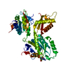
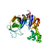
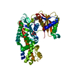
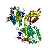
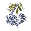
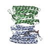
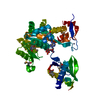
| Title | Crystal structure of human DUSP5 | ||||||
 Components Components | Dual specificity protein phosphatase 5 | ||||||
 Keywords Keywords |  HYDROLASE / alpha/beta HYDROLASE / alpha/beta | ||||||
| Function / homology |  Function and homology information Function and homology information: / protein serine/threonine phosphatase activity => GO:0004722 / protein serine/threonine phosphatase activity => GO:0004722 / : / protein tyrosine/threonine phosphatase activity / MAP kinase tyrosine/serine/threonine phosphatase activity / endoderm formation / peptidyl-threonine dephosphorylation / protein tyrosine/serine/threonine phosphatase activity / RAF-independent MAPK1/3 activation ...: / protein serine/threonine phosphatase activity => GO:0004722 / protein serine/threonine phosphatase activity => GO:0004722 / : / protein tyrosine/threonine phosphatase activity / MAP kinase tyrosine/serine/threonine phosphatase activity / endoderm formation / peptidyl-threonine dephosphorylation / protein tyrosine/serine/threonine phosphatase activity / RAF-independent MAPK1/3 activation / mitogen-activated protein kinase binding / protein-serine/threonine phosphatase / phosphatase activity / phosphoprotein phosphatase activity / peptidyl-tyrosine dephosphorylation / dephosphorylation / protein dephosphorylation / protein-tyrosine-phosphatase / protein tyrosine phosphatase activity / Negative regulation of MAPK pathway / MAPK cascade / nucleoplasm / nucleus / cytoplasmSimilarity search - Function | ||||||
| Biological species |  Homo sapiens (human) Homo sapiens (human) | ||||||
| Method | X-RAY DIFFRACTION / SYNCHROTRON / MOLECULAR REPLACEMENT / Resolution: 2.7 Å | ||||||
 Authors Authors | Kim, S.J. / Ryu, S.E. | ||||||
 Citation Citation | Journal: Proteins / Year: 2007 Title: Crystal structure of the catalytic domain of human DUSP5, a dual specificity MAP kinase protein phosphatase Authors: Jeong, D.G. / Cho, Y.H. / Yoon, T.S. / Kim, J.H. / Ryu, S.E. / Kim, S.J. | ||||||
| History |
|
- Structure visualization
| Structure viewer | Molecule: MolmilJmol/JSmol |
|---|
- Downloads & links
-Download
| PDBx/mmCIF format | 2g6z.cif.gz | 95.6 KB | Display | PDBx/mmCIF format |
|---|---|---|---|---|
| PDB format | pdb2g6z.ent.gz | 78 KB | Display | PDB format |
| PDBx/mmJSON format | 2g6z.json.gz | Tree view | PDBx/mmJSON format | |
| Others |  Other downloads Other downloads |
-Validation report
| Arichive directory | https://data.pdbj.org/pub/pdb/validation_reports/g6/2g6zftp://data.pdbj.org/pub/pdb/validation_reports/g6/2g6z | HTTPS FTP |
|---|
-Related structure data
| Similar structure data |
|---|


-Links
 PDBj
PDBj


- Assembly
| Deposited unit | 
| ||||||||
|---|---|---|---|---|---|---|---|---|---|
| 1 |
| ||||||||
| Unit cell |
| ||||||||
| Components on special symmetry positions |
| ||||||||
| Details | The biological assembly is dimer |
-Components
| #1: Protein | Mass: 23159.467 Da / Num. of mol.: 3 / Fragment: residues 174-384 Source method: isolated from a genetically manipulated source Source: (gene. exp.) Homo sapiens (human) / Production host:  Escherichia coli (E. coli) Escherichia coli (E. coli)References: UniProt: Q16690, protein-tyrosine-phosphatase, protein-serine/threonine phosphatase#2: Chemical | ChemComp-SO4 / Sulfate  Mass: 96.063 Da / Num. of mol.: 6 / Source method: obtained synthetically / Formula: SO4 Mass: 96.063 Da / Num. of mol.: 6 / Source method: obtained synthetically / Formula: SO4#3: Water | ChemComp-HOH / | Water Mass: 18.015 Da / Num. of mol.: 39 / Source method: isolated from a natural source / Formula: H2O Mass: 18.015 Da / Num. of mol.: 39 / Source method: isolated from a natural source / Formula: H2O |
|---|
-Experimental details
-Experiment
| Experiment | Method: X-RAY DIFFRACTION / Number of used crystals: 1 |
|---|
- Sample preparation
| Crystal | Density Matthews: 2.55 Å3/Da / Density % sol: 51.83 % |
|---|---|
| Crystal grow | Temperature: 291 K / Method: vapor diffusion, hanging drop / pH: 6.5 Details: 0.1M Bis-Tris pH 6.5, 2.2M ammonium sulfate and 6% (v/v) PEG 400, VAPOR DIFFUSION, HANGING DROP, temperature 291K |
-Data collection
| Diffraction | Mean temperature: 100 K |
|---|---|
| Diffraction source | Source: SYNCHROTRON / Site: Photon Factory  / Beamline: BL-5A / Wavelength: 1 Å / Beamline: BL-5A / Wavelength: 1 Å |
| Detector | Type: ADSC QUANTUM 315 / Detector: CCD / Date: Nov 23, 2004 |
| Radiation | Monochromator: Mirror / Protocol: SINGLE WAVELENGTH / Monochromatic (M) / Laue (L): M / Scattering type: x-ray |
| Radiation wavelength | Wavelength: 1 Å / Relative weight: 1 |
| Reflection | Resolution: 2.7→40 Å / Num. all: 20822 / Num. obs: 20524 / % possible obs: 98.6 % / Observed criterion σ(F): 0 / Observed criterion σ(I): 0 |
| Reflection shell | Resolution: 2.7→2.85 Å / % possible all: 100 |
- Processing
| Software |
| ||||||||||||||||||||
|---|---|---|---|---|---|---|---|---|---|---|---|---|---|---|---|---|---|---|---|---|---|
| Refinement | Method to determine structure: MOLECULAR REPLACEMENT / Resolution: 2.7→40 Å / σ(F): 0 / Stereochemistry target values: Engh & Huber
| ||||||||||||||||||||
| Refinement step | Cycle: LAST / Resolution: 2.7→40 Å
| ||||||||||||||||||||
| Refine LS restraints |
|
