 Movie
Movie Controller

[English] 日本語
 Yorodumi
- PDB-2fwm: Crystal Structure of E. coli EntA, a 2,3-dihydrodihydroxy benzoat... -
+ Open data

- Basic information
| Entry | Database: PDB / ID: 2fwm | ||||||
|---|---|---|---|---|---|---|---|
| Title | Crystal Structure of E. coli EntA, a 2,3-dihydrodihydroxy benzoate dehydrogenase | ||||||
 Components Components | 2,3-dihydro-2,3-dihydroxybenzoate dehydrogenase | ||||||
 Keywords Keywords | OXIDOREDUCTASE / enterobactin / Rossmann Fold / chorismate metabolism / short-chain oxidoreductase / tetramer | ||||||
| Function / homology |  Function and homology information2,3-dihydro-2,3-dihydroxybenzoate dehydrogenase / 2,3-dihydro-2,3-dihydroxybenzoate dehydrogenase activity / enterobactin biosynthetic process / protein-containing complex / identical protein binding / cytosol Function and homology information2,3-dihydro-2,3-dihydroxybenzoate dehydrogenase / 2,3-dihydro-2,3-dihydroxybenzoate dehydrogenase activity / enterobactin biosynthetic process / protein-containing complex / identical protein binding / cytosolSimilarity search - Function | ||||||
| Biological species |  Escherichia coli (E. coli) Escherichia coli (E. coli) | ||||||
| Method | X-RAY DIFFRACTION / SYNCHROTRON / MAD / Resolution: 2 Å | ||||||
 Authors Authors | Gulick, A.M. / Duax, W.L. | ||||||
 Citation Citation | Journal: Acta Crystallogr.,Sect.D / Year: 2006 Title: Determination of the crystal structure of EntA, a 2,3-dihydro-2,3-dihydroxybenzoic acid dehydrogenase from Escherichia coli. Authors: Sundlov, J.A. / Garringer, J.A. / Carney, J.M. / Reger, A.S. / Drake, E.J. / Duax, W.L. / Gulick, A.M. | ||||||
| History |
| ||||||
| Remark 999 | SEQUENCE After removal of the tag with TEV protease, a Gly-His sequence remains upstream of Met at position 1 |
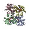
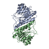
- Structure visualization
| Structure viewer | Molecule: MolmilJmol/JSmol |
|---|
- Downloads & links
-Download
| PDBx/mmCIF format | 2fwm.cif.gz | 53.8 KB | Display | PDBx/mmCIF format |
|---|---|---|---|---|
| PDB format | pdb2fwm.ent.gz | 38.2 KB | Display | PDB format |
| PDBx/mmJSON format | 2fwm.json.gz | Tree view | PDBx/mmJSON format | |
| Others |  Other downloads Other downloads |
-Validation report
| Arichive directory | https://data.pdbj.org/pub/pdb/validation_reports/fw/2fwmftp://data.pdbj.org/pub/pdb/validation_reports/fw/2fwm | HTTPS FTP |
|---|
-Related structure data
| Similar structure data |
|---|


-Links
 PDBj
PDBj


- Assembly
| Deposited unit | 
| ||||||||
|---|---|---|---|---|---|---|---|---|---|
| 1 | 
| ||||||||
| Unit cell |
| ||||||||
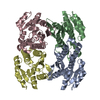
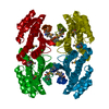
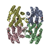
| Details | Tetramer formed around crystallographic 222 symmetry |
-Components
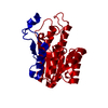
| #1: Protein | Mass: 26466.781 Da / Num. of mol.: 1 Source method: isolated from a genetically manipulated source Source: (gene. exp.) Escherichia coli (E. coli) / Strain: JM109 / Gene: entA / Plasmid: pET15b-TEV / Species (production host): Escherichia coli / Production host: Escherichia coli BL21(DE3) (bacteria) / Strain (production host): BL21(DE3)References: UniProt: P15047, 2,3-dihydro-2,3-dihydroxybenzoate dehydrogenase |
|---|---|
| #2: Water | ChemComp-HOH / Water Mass: 18.015 Da / Num. of mol.: 109 / Source method: isolated from a natural source / Formula: H2O Mass: 18.015 Da / Num. of mol.: 109 / Source method: isolated from a natural source / Formula: H2O |
-Experimental details
-Experiment
| Experiment | Method: X-RAY DIFFRACTION / Number of used crystals: 1 |
|---|
- Sample preparation
| Crystal | Density Matthews: 2.02 Å3/Da / Density % sol: 39.2 % |
|---|---|
| Crystal grow | Temperature: 277 K / pH: 5 Details: 2% PEG4000, 2% ethylene glycol, 300 mM AmSO4, 50 mM NaSuccinate, VAPOR DIFFUSION, HANGING DROP, temperature 277K, pH 5.00 |
-Data collection
| Diffraction | Mean temperature: 113 K |
|---|---|
| Diffraction source | Source: SYNCHROTRON / Site: CHESS  / Beamline: F2 / Wavelength: 0.9793 / Beamline: F2 / Wavelength: 0.9793 |
| Detector | Type: ADSC QUANTUM 4 / Detector: CCD / Date: Aug 2, 2005 |
| Radiation | Protocol: MAD / Monochromatic (M) / Laue (L): M / Scattering type: x-ray |
| Radiation wavelength | Wavelength: 0.9793 Å / Relative weight: 1 |
| Reflection | Resolution: 2→50 Å / Num. obs: 14485 / % possible obs: 95.4 % / Observed criterion σ(I): 0 / Redundancy: 4.3 % / Rmerge(I) obs: 0.059 |
| Reflection shell | Resolution: 2→2.07 Å / Redundancy: 3 % / Rmerge(I) obs: 0.298 / % possible all: 70 |
-Phasing
| Phasing | Method: MAD | |||||||||||||||||||||||||||||||||||||||||||||||||
|---|---|---|---|---|---|---|---|---|---|---|---|---|---|---|---|---|---|---|---|---|---|---|---|---|---|---|---|---|---|---|---|---|---|---|---|---|---|---|---|---|---|---|---|---|---|---|---|---|---|---|
| Phasing dm | FOM : 0.86 / FOM acentric: 0.86 / FOM centric: 0.83 / Reflection: 14387 / Reflection acentric: 12756 / Reflection centric: 1631 | |||||||||||||||||||||||||||||||||||||||||||||||||
| Phasing dm shell |
|
- Processing
| Software |
| ||||||||||||||||||||||||||||||||||||||||||||||||||||||||||||||||||||||||||||||||||||||||||
|---|---|---|---|---|---|---|---|---|---|---|---|---|---|---|---|---|---|---|---|---|---|---|---|---|---|---|---|---|---|---|---|---|---|---|---|---|---|---|---|---|---|---|---|---|---|---|---|---|---|---|---|---|---|---|---|---|---|---|---|---|---|---|---|---|---|---|---|---|---|---|---|---|---|---|---|---|---|---|---|---|---|---|---|---|---|---|---|---|---|---|---|
| Refinement | Method to determine structure: MAD / Resolution: 2→40 Å / Cor.coef. Fo:Fc: 0.946 / Cor.coef. Fo:Fc free: 0.935 / SU B: 3.454 / SU ML: 0.098 / Cross valid method: THROUGHOUT / σ(F): 0 / ESU R Free: 0.154 / Stereochemistry target values: Engh & Huber
| ||||||||||||||||||||||||||||||||||||||||||||||||||||||||||||||||||||||||||||||||||||||||||
| Solvent computation | Ion probe radii: 0.8 Å / Shrinkage radii: 0.8 Å / VDW probe radii: 1.2 Å / Solvent model: MASK | ||||||||||||||||||||||||||||||||||||||||||||||||||||||||||||||||||||||||||||||||||||||||||
| Displacement parameters | Biso mean: 21.48 Å2
| ||||||||||||||||||||||||||||||||||||||||||||||||||||||||||||||||||||||||||||||||||||||||||
| Refinement step | Cycle: LAST / Resolution: 2→40 Å
| ||||||||||||||||||||||||||||||||||||||||||||||||||||||||||||||||||||||||||||||||||||||||||
| Refine LS restraints |
| ||||||||||||||||||||||||||||||||||||||||||||||||||||||||||||||||||||||||||||||||||||||||||
| LS refinement shell | Resolution: 2→2.052 Å / Total num. of bins used: 20
|
