- PDB-2fop: The Crystal Structure of the N-terminal domain of HAUSP/USP7 comp... -
+
Open data
ID or keywords:
Loading...
-
Basic information
Entry
Database: PDB / ID: 2fop
Title
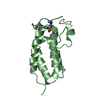
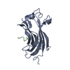
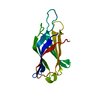
The Crystal Structure of the N-terminal domain of HAUSP/USP7 complexed with mdm2 peptide 147-150
Components
Ubiquitin carboxyl-terminal hydrolase 7
mdm2 peptide
Keywords
HYDROLASE / MATH Domain
Function / homology
Function and homology information
regulation of telomere capping / monoubiquitinated protein deubiquitination / regulation of retrograde transport, endosome to Golgi / cellular response to vitamin B1 / response to formaldehyde / response to water-immersion restraint stress / deubiquitinase activity / traversing start control point of mitotic cell cycle / negative regulation of intrinsic apoptotic signaling pathway by p53 class mediator / response to ether ...regulation of telomere capping / monoubiquitinated protein deubiquitination / regulation of retrograde transport, endosome to Golgi / cellular response to vitamin B1 / response to formaldehyde / response to water-immersion restraint stress / deubiquitinase activity / traversing start control point of mitotic cell cycle / negative regulation of intrinsic apoptotic signaling pathway by p53 class mediator / response to ether / negative regulation of signal transduction by p53 class mediator / fibroblast activation / atrial septum development / DNA alkylation repair / receptor serine/threonine kinase binding / Trafficking of AMPA receptors / positive regulation of vascular associated smooth muscle cell migration / regulation of DNA-binding transcription factor activity / negative regulation of gene expression via chromosomal CpG island methylation / peroxisome proliferator activated receptor binding / response to iron ion / negative regulation of protein processing / K48-linked deubiquitinase activity / SUMO transferase activity / response to steroid hormone / NEDD8 ligase activity / symbiont-mediated disruption of host cell PML body / AKT phosphorylates targets in the cytosol / atrioventricular valve morphogenesis / cellular response to peptide hormone stimulus / ventricular septum development / endocardial cushion morphogenesis / negative regulation of NF-kappaB transcription factor activity / positive regulation of muscle cell differentiation / SUMOylation of ubiquitinylation proteins / cellular response to alkaloid / blood vessel development / regulation of protein catabolic process / cardiac septum morphogenesis / protein deubiquitination / Constitutive Signaling by AKT1 E17K in Cancer / ligase activity / negative regulation of DNA damage response, signal transduction by p53 class mediator / response to magnesium ion / protein sumoylation / SUMOylation of transcription factors / protein localization to nucleus / negative regulation of proteasomal ubiquitin-dependent protein catabolic process / cellular response to UV-C / blood vessel remodeling / cellular response to estrogen stimulus / transcription-coupled nucleotide-excision repair / protein autoubiquitination / cellular response to actinomycin D / negative regulation of gluconeogenesis / ribonucleoprotein complex binding / negative regulation of TORC1 signaling / positive regulation of vascular associated smooth muscle cell proliferation / DNA damage response, signal transduction by p53 class mediator resulting in cell cycle arrest / NPAS4 regulates expression of target genes / transcription repressor complex / regulation of heart rate / Regulation of PTEN localization / positive regulation of mitotic cell cycle / Synthesis of active ubiquitin: roles of E1 and E2 enzymes / proteolysis involved in protein catabolic process / positive regulation of protein export from nucleus / regulation of signal transduction by p53 class mediator / response to cocaine / ubiquitin binding / Stabilization of p53 / Regulation of RUNX3 expression and activity / protein destabilization / RING-type E3 ubiquitin transferase / Signaling by ALK fusions and activated point mutants / establishment of protein localization / regulation of protein stability / Oncogene Induced Senescence / regulation of circadian rhythm / Regulation of TP53 Activity through Methylation / Transcription-Coupled Nucleotide Excision Repair (TC-NER) / cellular response to gamma radiation / Formation of TC-NER Pre-Incision Complex / response to toxic substance / cellular response to growth factor stimulus / PML body / cellular response to hydrogen peroxide / Dual incision in TC-NER / Gap-filling DNA repair synthesis and ligation in TC-NER / protein polyubiquitination / ubiquitin-protein transferase activity / endocytic vesicle membrane / ubiquitin protein ligase activity / disordered domain specific binding / Regulation of TP53 Degradation / rhythmic process / positive regulation of proteasomal ubiquitin-dependent protein catabolic process / p53 binding / negative regulation of neuron projection development / chromosome Similarity search - Function
Ubiquitin carboxyl-terminal hydrolase 7, ICP0-binding domain / ICP0-binding domain of Ubiquitin-specific protease 7 / Apoptosis, Tumor Necrosis Factor Receptor Associated Protein 2; Chain A / Apoptosis, Tumor Necrosis Factor Receptor Associated Protein 2; Chain A / Ubiquitin carboxyl-terminal hydrolase, C-terminal / Ubiquitin-specific protease C-terminal / MATH domain / E3 ubiquitin-protein ligase Mdm2 / MDM2, modified RING finger, HC subclass / MATH/TRAF domain ...Ubiquitin carboxyl-terminal hydrolase 7, ICP0-binding domain / ICP0-binding domain of Ubiquitin-specific protease 7 / Apoptosis, Tumor Necrosis Factor Receptor Associated Protein 2; Chain A / Apoptosis, Tumor Necrosis Factor Receptor Associated Protein 2; Chain A / Ubiquitin carboxyl-terminal hydrolase, C-terminal / Ubiquitin-specific protease C-terminal / MATH domain / E3 ubiquitin-protein ligase Mdm2 / MDM2, modified RING finger, HC subclass / MATH/TRAF domain / MATH/TRAF domain profile. / meprin and TRAF homology / TRAF-like / p53 negative regulator Mdm2/Mdm4 / SWIB/MDM2 domain / SWIB/MDM2 domain / SWIB/MDM2 domain profile. / SWIB/MDM2 domain superfamily / Ubiquitin specific protease (USP) domain signature 2. / Ubiquitin specific protease (USP) domain signature 1. / Ubiquitin specific protease, conserved site / Peptidase C19, ubiquitin carboxyl-terminal hydrolase / Ubiquitin carboxyl-terminal hydrolase / Ubiquitin specific protease domain / Ubiquitin specific protease (USP) domain profile. / Zn-finger in Ran binding protein and others / Zinc finger, C3HC4 type (RING finger) / Zinc finger RanBP2 type profile. / Zinc finger RanBP2-type signature. / Zinc finger, RanBP2-type superfamily / Zinc finger, RanBP2-type / Papain-like cysteine peptidase superfamily / Zinc finger RING-type profile. / Zinc finger, RING-type / Zinc finger, RING/FYVE/PHD-type / Sandwich / Mainly Beta Similarity search - Domain/homology
In the structure databanks used in Yorodumi, some data are registered as the other names, "COVID-19 virus" and "2019-nCoV". Here are the details of the virus and the list of structure data.
Jan 31, 2019. EMDB accession codes are about to change! (news from PDBe EMDB page)
EMDB accession codes are about to change! (news from PDBe EMDB page)
The allocation of 4 digits for EMDB accession codes will soon come to an end. Whilst these codes will remain in use, new EMDB accession codes will include an additional digit and will expand incrementally as the available range of codes is exhausted. The current 4-digit format prefixed with “EMD-” (i.e. EMD-XXXX) will advance to a 5-digit format (i.e. EMD-XXXXX), and so on. It is currently estimated that the 4-digit codes will be depleted around Spring 2019, at which point the 5-digit format will come into force.
The EM Navigator/Yorodumi systems omit the EMD- prefix.
Related info.:Q: What is EMD? / ID/Accession-code notation in Yorodumi/EM Navigator
Yorodumi is a browser for structure data from EMDB, PDB, SASBDB, etc.
This page is also the successor to EM Navigator detail page, and also detail information page/front-end page for Omokage search.
The word "yorodu" (or yorozu) is an old Japanese word meaning "ten thousand". "mi" (miru) is to see.
Related info.:EMDB / PDB / SASBDB / Comparison of 3 databanks / Yorodumi Search / Aug 31, 2016. New EM Navigator & Yorodumi / Yorodumi Papers / Jmol/JSmol / Function and homology information / Changes in new EM Navigator and Yorodumi
 Movie
Movie Controller

 Yorodumi
 Open data
 Basic information
 Components
 Keywords
 HYDROLASE /
  Function and homology information
 Authors
 Citation
 Structure visualization
 Downloads & links
 Other downloads


 PDBj
PDBj


 Assembly
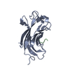

 Mass: 18.015 Da / Num. of mol.: 138 / Source method: isolated from a natural source / Formula: H2O
 Sample preparation
 Processing