 Movie
Movie Controller

[English] 日本語
 Yorodumi
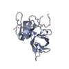
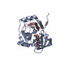
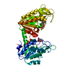
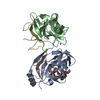
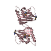
- PDB-2fm2: HCV NS3-4A protease domain complexed with a ketoamide inhibitor, ... -
+ Open data

- Basic information
| Entry | Database: PDB / ID: 2fm2 | ||||||
|---|---|---|---|---|---|---|---|
| Title | HCV NS3-4A protease domain complexed with a ketoamide inhibitor, SCH446211 | ||||||
 Components Components |
| ||||||
 Keywords Keywords |  HYDROLASE / Hepatitis C Virus / HCV / NS3/4A protease / ketoamide inhibitor HYDROLASE / Hepatitis C Virus / HCV / NS3/4A protease / ketoamide inhibitor | ||||||
| Function / homology |  Function and homology information Function and homology informationhost cell mitochondrial membrane / host cell lipid droplet / symbiont-mediated suppression of host TRAF-mediated signal transduction / transformation of host cell by virus / symbiont-mediated perturbation of host cell cycle G1/S transition checkpoint / symbiont-mediated suppression of host JAK-STAT cascade via inhibition of STAT1 activity / symbiont-mediated suppression of host cytoplasmic pattern recognition receptor signaling pathway via inhibition of MAVS activity / ribonucleoside triphosphate phosphatase activity / lipid droplet / protein complex oligomerization ...host cell mitochondrial membrane / host cell lipid droplet / symbiont-mediated suppression of host TRAF-mediated signal transduction / transformation of host cell by virus / symbiont-mediated perturbation of host cell cycle G1/S transition checkpoint / symbiont-mediated suppression of host JAK-STAT cascade via inhibition of STAT1 activity / symbiont-mediated suppression of host cytoplasmic pattern recognition receptor signaling pathway via inhibition of MAVS activity / ribonucleoside triphosphate phosphatase activity / lipid droplet / protein complex oligomerization / monoatomic ion channel activity / viral nucleocapsid / clathrin-dependent endocytosis of virus by host cell / RNA helicase activity / host cell perinuclear region of cytoplasm / host cell endoplasmic reticulum membrane / symbiont-mediated suppression of host type I interferon-mediated signaling pathway / ribonucleoprotein complex / induction by virus of host autophagy / virus-mediated perturbation of host defense response / viral RNA genome replication / cysteine-type endopeptidase activity / RNA-dependent RNA polymerase activity / serine-type endopeptidase activity / fusion of virus membrane with host endosome membrane / viral envelope / virion attachment to host cell / host cell plasma membrane / virion membrane / structural molecule activity / proteolysis / RNA binding / zinc ion binding / ATP binding / plasma membrane / cytoplasmSimilarity search - Function | ||||||
| Biological species |  Hepatitis C virus Hepatitis C virus | ||||||
| Method | X-RAY DIFFRACTION / FOURIER SYNTHESIS / Resolution: 2.7 Å | ||||||
 Authors Authors | Yi, M. / Tong, X. / Skelton, A. / Chase, R. / Chen, T. / Prongay, A. / Bogen, S.L. / Saksena, A.K. / Njoroge, F.G. / Veselenak, R.L. ...Yi, M. / Tong, X. / Skelton, A. / Chase, R. / Chen, T. / Prongay, A. / Bogen, S.L. / Saksena, A.K. / Njoroge, F.G. / Veselenak, R.L. / Pyles, R.B. / Bourne, N. / Malcolm, B.A. / Lemon, S.M. | ||||||
 Citation Citation | Journal: J.Biol.Chem. / Year: 2006 Title: Mutations conferring resistance to SCH6, a novel hepatitis C virus NS3/4A protease inhibitor. Reduced RNA replication fitness and partial rescue by second-site mutations Authors: Yi, M. / Tong, X. / Skelton, A. / Chase, R. / Chen, T. / Prongay, A. / Bogen, S.L. / Saksena, A.K. / Njoroge, F.G. / Veselenak, R.L. / Pyles, R.B. / Bourne, N. / Malcolm, B.A. / Lemon, S.M. | ||||||
| History |
| ||||||
| Remark 300 | BIOMOLECULE: 1 THIS ENTRY CONTAINS THE CRYSTALLOGRAPHIC ASYMMETRIC UNIT WHICH CONSISTS OF 4 CHAIN(S) ...BIOMOLECULE: 1 THIS ENTRY CONTAINS THE CRYSTALLOGRAPHIC ASYMMETRIC UNIT WHICH CONSISTS OF 4 CHAIN(S).THE ASYMMETRIC UNIT CONTAINS A DIMER OF THE PROTEASE DOMAIN WHICH IS THE N-TERMINAL 181 RESIDUES OF THE NS3 PROTEIN (631 RESIDUES) THAT CONTAINS BOTH THE PROTEASE DOMAIN AND THE HELICASE DOMAIN. BIOLOGICALLY THE PROTEASE DOMAIN FUNCTIONS AS PART OF THE LARGER (631 RESIDUE) PROTEIN. THE LARGER PROTEIN HAS NOT BEEN SHOWN TO EXIST AS A DIMER. THE SMALLER PROTEASE DOMAIN (RESIDUES 1-181) THAT HAVE BEEN EXPRESSED WITH AN N-TERMINAL T7 EPITOPE TAG AND A C-TERMINAL HIS TAG, EXISTS AS A DIMER IN THE ASYMMETRIC UNIT. IT IS UNKNOWN WHETHER THIS PROTEIN DOMAIN IS FUNCTIONAL AS A MONOMER OR A DIMER. |
- Structure visualization
| Structure viewer | Molecule: MolmilJmol/JSmol |
|---|
- Downloads & links
-Download
| PDBx/mmCIF format | 2fm2.cif.gz | 89.4 KB | Display | PDBx/mmCIF format |
|---|---|---|---|---|
| PDB format | pdb2fm2.ent.gz | 66.3 KB | Display | PDB format |
| PDBx/mmJSON format | 2fm2.json.gz | Tree view | PDBx/mmJSON format | |
| Others |  Other downloads Other downloads |
-Validation report
| Arichive directory | https://data.pdbj.org/pub/pdb/validation_reports/fm/2fm2ftp://data.pdbj.org/pub/pdb/validation_reports/fm/2fm2 | HTTPS FTP |
|---|
-Related structure data
| Related structure data |  139aS S: Starting model for refinement |
|---|---|
| Similar structure data |


-Links
 PDBj
PDBj


- Assembly
| Deposited unit | 
| ||||||||
|---|---|---|---|---|---|---|---|---|---|
| 1 |
| ||||||||
| Unit cell |
| ||||||||
| Details | The asymmetric unit contains a dimer of the protease domain which is the N-terminal 181 residues of the NS3 protein (631 residues) that contains both the protease domain and the helicase domain. Biological the protease domain functions as part of the larger (631 residue) protein. The larger protein has not been shown to exist as a dimer. The smaller protease domain (residues 1-181) that have been expressed with an N-terminal T7 epitope tag and a C-terminal His Tag, exists as a dimer in the asymmetric unit. This protein domain is believed to be functional as either a monomer or a dimer. |
-Components
-Protein / Protein/peptide , 2 types, 4 molecules ACBD
| #1: Protein | Mass: 21233.225 Da / Num. of mol.: 2 / Fragment: protease domain Source method: isolated from a genetically manipulated source Source: (gene. exp.) Hepatitis C virus / Genus: Hepacivirus / Strain: H1A / Gene: NS polyprotein / Plasmid: pET-3A / Production host:  Escherichia coli (E. coli) / Strain (production host): BLR(DE3) / References: GenBank: 28921568, UniProt: Q9ELS8*PLUS Escherichia coli (E. coli) / Strain (production host): BLR(DE3) / References: GenBank: 28921568, UniProt: Q9ELS8*PLUS#2: Protein/peptide | Mass: 2394.039 Da / Num. of mol.: 2 Fragment: residues 24-39 with 2 LYS at both C and N-terminal Source method: obtained synthetically Details: Peptides were synthesized using Fmoc solid-phase chemistry on an ABI 431 synthesizer References: UniProt: Q9ELS8*PLUS |
|---|
-Non-polymers , 4 types, 158 molecules 


| #3: Chemical |  Mass: 65.409 Da / Num. of mol.: 2 / Source method: obtained synthetically / Formula: Zn Mass: 65.409 Da / Num. of mol.: 2 / Source method: obtained synthetically / Formula: Zn#4: Chemical | ChemComp-BME / | 2-Mercaptoethanol Mass: 78.133 Da / Num. of mol.: 1 / Source method: obtained synthetically / Formula: C2H6OS Mass: 78.133 Da / Num. of mol.: 1 / Source method: obtained synthetically / Formula: C2H6OS#5: Chemical | ChemComp-3BC / | Butyl group Mass: 724.887 Da / Num. of mol.: 1 / Source method: obtained synthetically / Formula: C38H56N6O8 Mass: 724.887 Da / Num. of mol.: 1 / Source method: obtained synthetically / Formula: C38H56N6O8#6: Water | ChemComp-HOH / | WaterMass: 18.015 Da / Num. of mol.: 154 / Source method: isolated from a natural source / Formula: H2O |
|---|
-Experimental details
-Experiment
| Experiment | Method: X-RAY DIFFRACTION / Number of used crystals: 1 |
|---|
- Sample preparation
| Crystal | Density Matthews: 3.88 Å3/Da / Density % sol: 68.31 % |
|---|---|
| Crystal grow | Temperature: 285 K / Method: vapor diffusion, hanging drop / pH: 5.7 Details: Crystallization was performed by the vapor diffusion method using hanging drops (4 uL protein solution mixed with 4 uL (0.75 - 1.00)M NaCl - 0.1M MES - 0.1M Na/K PO4, pH 5.6 - 5.8) suspended ...Details: Crystallization was performed by the vapor diffusion method using hanging drops (4 uL protein solution mixed with 4 uL (0.75 - 1.00)M NaCl - 0.1M MES - 0.1M Na/K PO4, pH 5.6 - 5.8) suspended over 1mL reservoir solutions of (1.25 - 1.50)M NaCl - 0.1M MES - 0.1M Na/K PO4 - 5mM-mercaptoethanol, pH 5.6-5.8. The trays were set at 4C for 5-7 days to control nucleation, followed by incubation for 3 weeks at 12C to maximize crystal growth. , pH 5.7, VAPOR DIFFUSION, HANGING DROP, temperature 285K |
-Data collection
| Diffraction | Mean temperature: 95 K |
|---|---|
| Diffraction source | Source: ROTATING ANODE / Type: RIGAKU RUH3R / Wavelength: 1.5418 Å |
| Detector | Type: RIGAKU RAXIS IV / Detector: IMAGE PLATE / Date: Apr 14, 2005 / Details: MULTILAYER X-RAY FOCUSING OPTICS |
| Radiation | Protocol: SINGLE WAVELENGTH / Monochromatic (M) / Laue (L): M / Scattering type: x-ray |
| Radiation wavelength | Wavelength: 1.5418 Å / Relative weight: 1 |
| Reflection | Resolution: 2.7→50 Å / Num. all: 19889 / Num. obs: 19511 / % possible obs: 98.1 % / Observed criterion σ(F): 1.7 / Observed criterion σ(I): 3 / Redundancy: 4 % / Biso Wilson estimate: 32.938 Å2 / Rsym value: 0.088 / Net I/σ(I): 13.6 |
| Reflection shell | Resolution: 2.7→2.78 Å / Redundancy: 2.6 % / Mean I/σ(I) obs: 3 / Num. unique all: 1636 / Rsym value: 0.323 / % possible all: 84.7 |
- Processing
| Software |
| |||||||||||||||||||||||||
|---|---|---|---|---|---|---|---|---|---|---|---|---|---|---|---|---|---|---|---|---|---|---|---|---|---|---|
| Refinement | Method to determine structure: FOURIER SYNTHESIS Starting model: HCV NS3(139A)/4A protease domain structure Resolution: 2.7→8 Å / σ(F): 1.7 / σ(I): 3 / Stereochemistry target values: Engh & Huber
| |||||||||||||||||||||||||
| Refinement step | Cycle: LAST / Resolution: 2.7→8 Å
| |||||||||||||||||||||||||
| Refine LS restraints |
| |||||||||||||||||||||||||
| LS refinement shell | Resolution: 2.7→2.82 Å
|
