 Movie
Movie Controller


+ Open data

- Basic information
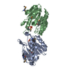
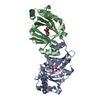
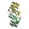
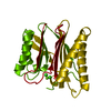
| Entry | Database: PDB / ID: 2fjt | ||||||
|---|---|---|---|---|---|---|---|
| Title | Adenylyl cyclase class iv from Yersinia pestis | ||||||
 Components Components | Adenylyl cyclase class IV | ||||||
 Keywords Keywords |  LYASE / CYCLASE / beta barrel / dimer LYASE / CYCLASE / beta barrel / dimer | ||||||
| Function / homology |  Function and homology informationadenylate cyclase / lyase activity / nucleotide binding / metal ion binding Function and homology informationadenylate cyclase / lyase activity / nucleotide binding / metal ion bindingSimilarity search - Function | ||||||
| Biological species |   Yersinia pestis (bacteria) Yersinia pestis (bacteria) | ||||||
| Method | X-RAY DIFFRACTION / MOLECULAR REPLACEMENT / Resolution: 1.901 Å | ||||||
 Authors Authors | Gallagher, D.T. / Smith, N.N. / Kim, S.-K. / Reddy, P.T. / Robinson, H. / Heroux, A. | ||||||
 Citation Citation | Journal: J.Mol.Biol. / Year: 2006 Title: Structure of the class IV adenylyl cyclase reveals a novel fold Authors: Gallagher, D.T. / Smith, N.N. / Kim, S.-K. / Heroux, A. / Robinson, H. / Reddy, P.T. #1: Journal: Acta Crystallogr.,Sect.F / Year: 2006Title: Crystallization of the class IV adenylyl cyclase from Yersinia pestis Authors: Smith, N.N. / Kim, S.-K. / Reddy, P.T. / Gallagher, D.T. | ||||||
| History |
| ||||||
| Remark 300 | BIOMOLECULE: 1 QUATERNARY STRUCTURE FOR THIS ENTRY: DIMER THIS ENTRY CONTAINS THE CRYSTALLOGRAPHIC ... BIOMOLECULE: 1 QUATERNARY STRUCTURE FOR THIS ENTRY: DIMER THIS ENTRY CONTAINS THE CRYSTALLOGRAPHIC ASYMMETRIC UNIT WHICH CONSISTS OF 2 CHAINS (1 BIOLOGICAL DIMER). SEE REMARK 350 FOR INFORMATION ON GENERATING THE BIOLOGICAL MOLECULE(S). |
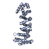
- Structure visualization
| Structure viewer | Molecule: MolmilJmol/JSmol |
|---|
- Downloads & links
-Download
| PDBx/mmCIF format | 2fjt.cif.gz | 89.2 KB | Display | PDBx/mmCIF format |
|---|---|---|---|---|
| PDB format | pdb2fjt.ent.gz | 66.8 KB | Display | PDB format |
| PDBx/mmJSON format | 2fjt.json.gz | Tree view | PDBx/mmJSON format | |
| Others |  Other downloads Other downloads |
-Validation report
| Arichive directory | https://data.pdbj.org/pub/pdb/validation_reports/fj/2fjtftp://data.pdbj.org/pub/pdb/validation_reports/fj/2fjt | HTTPS FTP |
|---|
-Related structure data
| Similar structure data |
|---|


-Links
 PDBj
PDBj


- Assembly
| Deposited unit | 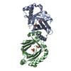
| ||||||||
|---|---|---|---|---|---|---|---|---|---|
| 1 |
| ||||||||
| Unit cell |
| ||||||||
| Details | the biol. assembly is a homodimer equal to the asymmetric unit. |
-Components
| #1: Protein | Mass: 20638.326 Da / Num. of mol.: 2 Source method: isolated from a genetically manipulated source Details: Triclinic form, pH 4.6 / Source: (gene. exp.) Yersinia pestis (bacteria) / Strain: KIM / Production host: Escherichia coli (E. coli)References: UniProt: Q7CH76, UniProt: A0A5P8YEL9*PLUS, adenylate cyclase#2: Chemical | Sulfate  Mass: 96.063 Da / Num. of mol.: 2 / Source method: obtained synthetically / Formula: SO4 Mass: 96.063 Da / Num. of mol.: 2 / Source method: obtained synthetically / Formula: SO4#3: Water | ChemComp-HOH / | Water Mass: 18.015 Da / Num. of mol.: 304 / Source method: isolated from a natural source / Formula: H2O Mass: 18.015 Da / Num. of mol.: 304 / Source method: isolated from a natural source / Formula: H2O |
|---|
-Experimental details
-Experiment
| Experiment | Method: X-RAY DIFFRACTION / Number of used crystals: 1 |
|---|
- Sample preparation
| Crystal | Density Matthews: 1.87 Å3/Da / Density % sol: 34.35 % |
|---|---|
| Crystal grow | Temperature: 297 K / Method: vapor diffusion, hanging drop / pH: 4.6 Details: 100 mM Ammonium sulfate, 100 mM Sodium acetate, 14% PEG 4000, pH 4.6, VAPOR DIFFUSION, HANGING DROP, temperature 297K |
-Data collection
| Diffraction source | Source: ROTATING ANODE / Type: RIGAKU / Wavelength: 1.5418 Å |
|---|---|
| Detector | Type: RIGAKU RAXIS IV / Detector: IMAGE PLATE / Date: Sep 20, 2004 |
| Radiation | Monochromator: mirrors / Protocol: SINGLE WAVELENGTH / Monochromatic (M) / Laue (L): M / Scattering type: x-ray |
| Radiation wavelength | Wavelength: 1.5418 Å / Relative weight: 1 |
| Reflection | Resolution: 1.9→20 Å / Num. all: 21019 / Num. obs: 20986 / % possible obs: 89.3 % / Observed criterion σ(F): 0 / Observed criterion σ(I): 0 / Redundancy: 2.04 % / Rmerge(I) obs: 0.026 / Net I/σ(I): 15.7 |
| Reflection shell | Resolution: 1.9→1.97 Å / Redundancy: 2.04 % / Rmerge(I) obs: 0.17 / Mean I/σ(I) obs: 3.9 / % possible all: 83.9 |
- Processing
| Software |
| ||||||||||||||||||||||||||||||||||||||||||||||||||||||||||||||||||||||||||||||||||||||||||
|---|---|---|---|---|---|---|---|---|---|---|---|---|---|---|---|---|---|---|---|---|---|---|---|---|---|---|---|---|---|---|---|---|---|---|---|---|---|---|---|---|---|---|---|---|---|---|---|---|---|---|---|---|---|---|---|---|---|---|---|---|---|---|---|---|---|---|---|---|---|---|---|---|---|---|---|---|---|---|---|---|---|---|---|---|---|---|---|---|---|---|---|
| Refinement | Method to determine structure: MOLECULAR REPLACEMENT Starting model: orthorhombic crystal form Resolution: 1.901→9.98 Å / Cor.coef. Fo:Fc: 0.954 / Cor.coef. Fo:Fc free: 0.926 / SU B: 5.679 / SU ML: 0.166 / Cross valid method: THROUGHOUT / σ(F): 0 / ESU R: 0.286 / ESU R Free: 0.221 / Stereochemistry target values: MAXIMUM LIKELIHOOD
| ||||||||||||||||||||||||||||||||||||||||||||||||||||||||||||||||||||||||||||||||||||||||||
| Solvent computation | Ion probe radii: 0.8 Å / Shrinkage radii: 0.8 Å / VDW probe radii: 1.2 Å / Solvent model: MASK | ||||||||||||||||||||||||||||||||||||||||||||||||||||||||||||||||||||||||||||||||||||||||||
| Displacement parameters | Biso mean: 30.005 Å2
| ||||||||||||||||||||||||||||||||||||||||||||||||||||||||||||||||||||||||||||||||||||||||||
| Refine analyze |
| ||||||||||||||||||||||||||||||||||||||||||||||||||||||||||||||||||||||||||||||||||||||||||
| Refinement step | Cycle: LAST / Resolution: 1.901→9.98 Å
| ||||||||||||||||||||||||||||||||||||||||||||||||||||||||||||||||||||||||||||||||||||||||||
| Refine LS restraints |
| ||||||||||||||||||||||||||||||||||||||||||||||||||||||||||||||||||||||||||||||||||||||||||
| LS refinement shell | Resolution: 1.901→1.949 Å / Total num. of bins used: 20
|
