- PDB-2ffb: The crystal structure of the HlyB-NBD E631Q mutant in complex with ADP -
+
Open data
ID or keywords:
Loading...
-
Basic information
Entry
Database: PDB / ID: 2ffb
Title
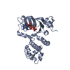
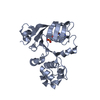
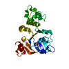
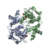
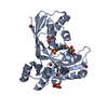
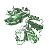
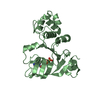
The crystal structure of the HlyB-NBD E631Q mutant in complex with ADP
Components
Alpha-hemolysin translocation ATP-binding protein hlyB
Keywords
TRANSPORT PROTEIN / ABC-transporter / ATPase
Function / homology
Function and homology information
protein secretion by the type I secretion system / type I protein secretion system complex / ATPase-coupled lipid transmembrane transporter activity / ABC-type transporter activity / peptidase activity / ATP hydrolysis activity / proteolysis / ATP binding / plasma membrane Similarity search - Function
ATPase, type I secretion system, HlyB / Peptidase C39-like A / Peptidase C39 family / Peptidase C39, bacteriocin processing / Peptidase family C39 domain profile. / Type 1 protein exporter / ABC transporter transmembrane region / ABC transporter type 1, transmembrane domain / ABC transporter integral membrane type-1 fused domain profile. / ABC transporter type 1, transmembrane domain superfamily ...ATPase, type I secretion system, HlyB / Peptidase C39-like A / Peptidase C39 family / Peptidase C39, bacteriocin processing / Peptidase family C39 domain profile. / Type 1 protein exporter / ABC transporter transmembrane region / ABC transporter type 1, transmembrane domain / ABC transporter integral membrane type-1 fused domain profile. / ABC transporter type 1, transmembrane domain superfamily / ABC transporter-like, conserved site / ABC transporters family signature. / ABC transporter / ABC transporter-like, ATP-binding domain / ATP-binding cassette, ABC transporter-type domain profile. / P-loop containing nucleotide triphosphate hydrolases / ATPases associated with a variety of cellular activities / AAA+ ATPase domain / P-loop containing nucleoside triphosphate hydrolase / Rossmann fold / 3-Layer(aba) Sandwich / Alpha Beta Similarity search - Domain/homology
In the structure databanks used in Yorodumi, some data are registered as the other names, "COVID-19 virus" and "2019-nCoV". Here are the details of the virus and the list of structure data.
Jan 31, 2019. EMDB accession codes are about to change! (news from PDBe EMDB page)
EMDB accession codes are about to change! (news from PDBe EMDB page)
The allocation of 4 digits for EMDB accession codes will soon come to an end. Whilst these codes will remain in use, new EMDB accession codes will include an additional digit and will expand incrementally as the available range of codes is exhausted. The current 4-digit format prefixed with “EMD-” (i.e. EMD-XXXX) will advance to a 5-digit format (i.e. EMD-XXXXX), and so on. It is currently estimated that the 4-digit codes will be depleted around Spring 2019, at which point the 5-digit format will come into force.
The EM Navigator/Yorodumi systems omit the EMD- prefix.
Related info.:Q: What is EMD? / ID/Accession-code notation in Yorodumi/EM Navigator
Yorodumi is a browser for structure data from EMDB, PDB, SASBDB, etc.
This page is also the successor to EM Navigator detail page, and also detail information page/front-end page for Omokage search.
The word "yorodu" (or yorozu) is an old Japanese word meaning "ten thousand". "mi" (miru) is to see.
Related info.:EMDB / PDB / SASBDB / Comparison of 3 databanks / Yorodumi Search / Aug 31, 2016. New EM Navigator & Yorodumi / Yorodumi Papers / Jmol/JSmol / Function and homology information / Changes in new EM Navigator and Yorodumi
 Movie
Movie Controller

 Yorodumi
 Open data
 Basic information
 Components
 Keywords
 TRANSPORT PROTEIN /
  Function and homology information
 Authors
 Citation
 Structure visualization
 Downloads & links
 Other downloads


 PDBj
PDBj

 Assembly

 Mass: 427.201 Da / Num. of mol.: 1 / Source method: obtained synthetically / Formula: C10H15N5O10P2 / Comment: ADP, energy-carrying molecule*YM
 Mass: 18.015 Da / Num. of mol.: 128 / Source method: isolated from a natural source / Formula: H2O
 Sample preparation
 / Beamline: BW6 / Wavelength: 1.05 Å
 Processing