 Movie
Movie Controller


+ Open data

- Basic information
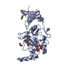
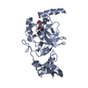
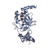
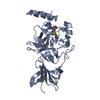
| Entry | Database: PDB / ID: 2f69 | ||||||
|---|---|---|---|---|---|---|---|
| Title | Ternary complex of SET7/9 bound to AdoHcy and a TAF10 peptide | ||||||
 Components Components |
| ||||||
 Keywords Keywords |  TRANSFERASE / SET domain / protein lysine methyltransferase / enzyme-peptide-AdoHcy complex TRANSFERASE / SET domain / protein lysine methyltransferase / enzyme-peptide-AdoHcy complex | ||||||
| Function / homology |  Function and homology information Function and homology informationSAGA complex assembly / lateral mesodermal cell differentiation / allantois development / peptidyl-lysine monomethylation / peptidyl-lysine dimethylation / transcription factor TFTC complex / [histone H3]-lysine4 N-methyltransferase / histone H3K4 monomethyltransferase activity / hepatocyte differentiation / protein-lysine N-methyltransferase activity ...SAGA complex assembly / lateral mesodermal cell differentiation / allantois development / peptidyl-lysine monomethylation / peptidyl-lysine dimethylation / transcription factor TFTC complex / [histone H3]-lysine4 N-methyltransferase / histone H3K4 monomethyltransferase activity / hepatocyte differentiation / protein-lysine N-methyltransferase activity / RNA polymerase binding / histone H3 methyltransferase activity / SAGA complex / limb development / transcription preinitiation complex / histone methyltransferase activity / HIV Transcription Initiation / RNA Polymerase II HIV Promoter Escape / Transcription of the HIV genome / RNA Polymerase II Promoter Escape / RNA Polymerase II Transcription Pre-Initiation And Promoter Opening / RNA Polymerase II Transcription Initiation / RNA Polymerase II Transcription Initiation And Promoter Clearance / regulation of RNA splicing / transcription factor TFIID complex / heterochromatin organization / RNA polymerase II general transcription initiation factor activity / positive regulation of transcription initiation by RNA polymerase II / embryonic placenta development / regulation of DNA repair / somitogenesis / RNA polymerase II preinitiation complex assembly / RNA Polymerase II Pre-transcription Events / male germ cell nucleus / DNA-templated transcription initiation / promoter-specific chromatin binding / nuclear estrogen receptor binding / transcription initiation at RNA polymerase II promoter / mRNA transcription by RNA polymerase II / multicellular organism growth / G1/S transition of mitotic cell cycle / PKMTs methylate histone lysines / p53 binding / chromosome / chromatin organization / HATs acetylate histones / response to ethanol / Regulation of TP53 Activity through Phosphorylation / transcription by RNA polymerase II / Ub-specific processing proteases / chromatin remodeling / apoptotic process / DNA damage response / chromatin binding / regulation of DNA-templated transcription / nucleolus / regulation of transcription by RNA polymerase II / perinuclear region of cytoplasm / positive regulation of DNA-templated transcription / enzyme binding / DNA binding / nucleoplasm / identical protein binding / nucleus / cytoplasmSimilarity search - Function | ||||||
| Biological species |  Homo sapiens (human) Homo sapiens (human) | ||||||
| Method | X-RAY DIFFRACTION / SYNCHROTRON / MOLECULAR REPLACEMENT / Resolution: 1.3 Å | ||||||
 Authors Authors | Couture, J.-F. / Collazo, E. / Hauk, G. / Trievel, R.C. | ||||||
 Citation Citation | Journal: Nat.Struct.Mol.Biol. / Year: 2006 Title: Structural basis for the methylation site specificity of SET7/9 Authors: Couture, J.-F. / Collazo, E. / Hauk, G. / Trievel, R.C. | ||||||
| History |
|
- Structure visualization
| Structure viewer | Molecule: MolmilJmol/JSmol |
|---|
- Downloads & links
-Download
| PDBx/mmCIF format | 2f69.cif.gz | 139.7 KB | Display | PDBx/mmCIF format |
|---|---|---|---|---|
| PDB format | pdb2f69.ent.gz | 106.2 KB | Display | PDB format |
| PDBx/mmJSON format | 2f69.json.gz | Tree view | PDBx/mmJSON format | |
| Others |  Other downloads Other downloads |
-Validation report
| Arichive directory | https://data.pdbj.org/pub/pdb/validation_reports/f6/2f69ftp://data.pdbj.org/pub/pdb/validation_reports/f6/2f69 | HTTPS FTP |
|---|
-Related structure data
| Related structure data | 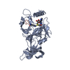 1o9sS S: Starting model for refinement |
|---|---|
| Similar structure data |


-Links
 PDBj
PDBj

- Assembly
| Deposited unit | 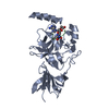
| ||||||||
|---|---|---|---|---|---|---|---|---|---|
| 1 |
| ||||||||
| Unit cell |
| ||||||||
| Components on special symmetry positions |
|
-Components
| #1: Protein | Histone methyltransferase / Histone H3-K4 methyltransferase / H3-K4-HMTase / SET domain-containing protein 7 / Set9 / SET7/9 Mass: 29043.330 Da / Num. of mol.: 1 Source method: isolated from a genetically manipulated source Source: (gene. exp.) Homo sapiens (human) / Gene: SET7, KIAA1717 / Plasmid: pHIS2 / Species (production host): Escherichia coli / Production host:  Escherichia coli BL21 (bacteria) / Strain (production host): BL21 Escherichia coli BL21 (bacteria) / Strain (production host): BL21References: UniProt: Q8WTS6, histone-lysine N-methyltransferase |
|---|---|
| #2: Protein/peptide | Mass: 1268.484 Da / Num. of mol.: 1 / Source method: obtained synthetically / Details: Synthetic Human TAF10 peptide / References: UniProt: Q12962*PLUS |
| #3: Chemical | ChemComp-SAH / S-Adenosyl-L-homocysteine  Type: L-peptide linking / Mass: 384.411 Da / Num. of mol.: 1 / Source method: obtained synthetically / Formula: C14H20N6O5S Type: L-peptide linking / Mass: 384.411 Da / Num. of mol.: 1 / Source method: obtained synthetically / Formula: C14H20N6O5S |
| #4: Water | ChemComp-HOH / Water Mass: 18.015 Da / Num. of mol.: 472 / Source method: isolated from a natural source / Formula: H2O Mass: 18.015 Da / Num. of mol.: 472 / Source method: isolated from a natural source / Formula: H2O |
-Experimental details
-Experiment
| Experiment | Method: X-RAY DIFFRACTION / Number of used crystals: 1 |
|---|
- Sample preparation
| Crystal | Density Matthews: 3.19 Å3/Da / Density % sol: 61.49 % |
|---|---|
| Crystal grow | Temperature: 293 K / Method: vapor diffusion, hanging drop / pH: 6.5 Details: 1.89-2.04 M (NH4)2SO4, 0.1 M Bis TRIS pH 6.5, VAPOR DIFFUSION, HANGING DROP, temperature 293.0K |
-Data collection
| Diffraction | Mean temperature: 95 K |
|---|---|
| Diffraction source | Source: SYNCHROTRON / Site: APS  / Beamline: 17-ID / Wavelength: 0.96863 Å / Beamline: 17-ID / Wavelength: 0.96863 Å |
| Detector | Type: ADSC QUANTUM 210 / Detector: CCD / Date: Aug 13, 2005 |
| Radiation | Monochromator: Si 111 / Protocol: SINGLE WAVELENGTH / Monochromatic (M) / Laue (L): M / Scattering type: x-ray |
| Radiation wavelength | Wavelength: 0.96863 Å / Relative weight: 1 |
| Reflection | Resolution: 1.3→29.14 Å / Num. all: 94528 / Num. obs: 94528 / % possible obs: 100 % / Observed criterion σ(I): -3 / Redundancy: 11.26 % / Rmerge(I) obs: 0.06 / Net I/σ(I): 17 |
| Reflection shell | Resolution: 1.3→1.35 Å / Redundancy: 9.32 % / Rmerge(I) obs: 0.505 / Mean I/σ(I) obs: 3.9 / Num. unique all: 9356 / % possible all: 100 |
- Processing
| Software |
| ||||||||||||||||||||||||||||||||||||||||||||||||||||||||||||||||||||||||||||||||||||||||||||||||||||||||||||||||||||||||||||||||||||||||||||||||||||||||||||||||||||||||||
|---|---|---|---|---|---|---|---|---|---|---|---|---|---|---|---|---|---|---|---|---|---|---|---|---|---|---|---|---|---|---|---|---|---|---|---|---|---|---|---|---|---|---|---|---|---|---|---|---|---|---|---|---|---|---|---|---|---|---|---|---|---|---|---|---|---|---|---|---|---|---|---|---|---|---|---|---|---|---|---|---|---|---|---|---|---|---|---|---|---|---|---|---|---|---|---|---|---|---|---|---|---|---|---|---|---|---|---|---|---|---|---|---|---|---|---|---|---|---|---|---|---|---|---|---|---|---|---|---|---|---|---|---|---|---|---|---|---|---|---|---|---|---|---|---|---|---|---|---|---|---|---|---|---|---|---|---|---|---|---|---|---|---|---|---|---|---|---|---|---|---|---|
| Refinement | Method to determine structure: MOLECULAR REPLACEMENT Starting model: PDB ENTRY 1O9S Resolution: 1.3→29.14 Å / Cor.coef. Fo:Fc: 0.976 / Cor.coef. Fo:Fc free: 0.97 / SU B: 1.241 / SU ML: 0.023 / Cross valid method: THROUGHOUT / σ(F): 3 / ESU R: 0.038 / ESU R Free: 0.039 / Stereochemistry target values: MAXIMUM LIKELIHOOD / Details: HYDROGENS HAVE BEEN ADDED IN THE RIDING POSITIONS
| ||||||||||||||||||||||||||||||||||||||||||||||||||||||||||||||||||||||||||||||||||||||||||||||||||||||||||||||||||||||||||||||||||||||||||||||||||||||||||||||||||||||||||
| Solvent computation | Ion probe radii: 0.8 Å / Shrinkage radii: 0.8 Å / VDW probe radii: 1.2 Å / Solvent model: MASK | ||||||||||||||||||||||||||||||||||||||||||||||||||||||||||||||||||||||||||||||||||||||||||||||||||||||||||||||||||||||||||||||||||||||||||||||||||||||||||||||||||||||||||
| Displacement parameters | Biso mean: 24.263 Å2
| ||||||||||||||||||||||||||||||||||||||||||||||||||||||||||||||||||||||||||||||||||||||||||||||||||||||||||||||||||||||||||||||||||||||||||||||||||||||||||||||||||||||||||
| Refinement step | Cycle: LAST / Resolution: 1.3→29.14 Å
| ||||||||||||||||||||||||||||||||||||||||||||||||||||||||||||||||||||||||||||||||||||||||||||||||||||||||||||||||||||||||||||||||||||||||||||||||||||||||||||||||||||||||||
| Refine LS restraints |
| ||||||||||||||||||||||||||||||||||||||||||||||||||||||||||||||||||||||||||||||||||||||||||||||||||||||||||||||||||||||||||||||||||||||||||||||||||||||||||||||||||||||||||
| LS refinement shell | Resolution: 1.3→1.334 Å / Total num. of bins used: 20
|
