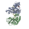
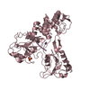
Entry Database : PDB / ID : 2f55Title Two hepatitis c virus ns3 helicase domains complexed with the same strand of dna 5'-D(P*(DU)P*(DU)P*(DU))-3'5'-D(P*(DU)P*(DU)P*(DU)P*(DU)P*(DU)P*(DU)P*(DU)P*(DU)P*(DU)P*(DU)P*(DU)P*(DU)P*(DU))-3'polyprotein Keywords / Function / homology Function Domain/homology Component
/ / / / / / / / / / / / / / / / / / / / / / / / / / / / / / / / / / / / / / / / / / / / / / / / / / / / / / / / / / / / / / / / / / / / / / / / / / / / / / / / / / / / / / / / / / / / / / / / / / / / / / / / / / / / / / / / / / Biological species Method / / Resolution : 3.3 Å Authors Lu, J.Z. / Jordan, J.B. / Sakon, J. Journal : J.Biol.Chem. / Year : 2006Title : Structural and biological identification of residues on the surface of NS3 helicase required for optimal replication of the hepatitis C virusAuthors : Mackintosh, S.G. / Lu, J.Z. / Jordan, J.B. / Harrison, M.K. / Sikora, B. / Sharma, S.D. / Cameron, C.E. / Raney, K.D. / Sakon, J. History Deposition Nov 25, 2005 Deposition site / Processing site Revision 1.0 Dec 6, 2005 Provider / Type Revision 1.1 May 1, 2008 Group Revision 1.2 Jul 13, 2011 Group Revision 1.3 Oct 18, 2017 Group / Category Revision 1.4 Aug 23, 2023 Group Data collection / Database references ... Data collection / Database references / Derived calculations / Refinement description Category chem_comp_atom / chem_comp_bond ... chem_comp_atom / chem_comp_bond / database_2 / pdbx_initial_refinement_model / struct_site Item _database_2.pdbx_DOI / _database_2.pdbx_database_accession ... _database_2.pdbx_DOI / _database_2.pdbx_database_accession / _struct_site.pdbx_auth_asym_id / _struct_site.pdbx_auth_comp_id / _struct_site.pdbx_auth_seq_id
Show all Show less
 Movie
Movie Controller

 Yorodumi
 Open data
 Basic information
 Components
 Keywords
 Function and homology information
 hepacivirin / host cell mitochondrial membrane / host cell lipid droplet / symbiont-mediated suppression of host TRAF-mediated signal transduction / transformation of host cell by virus / symbiont-mediated perturbation of host cell cycle G1/S transition checkpoint / symbiont-mediated suppression of host JAK-STAT cascade via inhibition of STAT1 activity / symbiont-mediated suppression of host cytoplasmic pattern recognition receptor signaling pathway via inhibition of MAVS activity /
  Hepatitis C virus
 Authors
 Citation
 Structure visualization
 Downloads & links
 Other downloads


 PDBj
PDBj


 Assembly
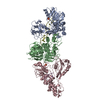


 Mass: 96.063 Da / Num. of mol.: 2 / Source method: obtained synthetically / Formula: SO4
 Mass: 18.015 Da / Num. of mol.: 62 / Source method: isolated from a natural source / Formula: H2O
 Sample preparation
 Processing