 Movie
Movie Controller


+ Open data

- Basic information
| Entry | Database: PDB / ID: 2ewt | ||||||
|---|---|---|---|---|---|---|---|
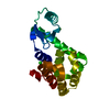
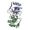
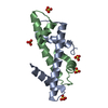
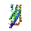
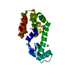
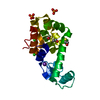
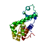
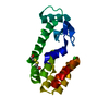
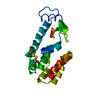
| Title | Crystal structure of the DNA-binding domain of BldD | ||||||
 Components Components | putative DNA-binding protein | ||||||
 Keywords Keywords | DNA BINDING PROTEIN / the DNA-binding domain of BldD | ||||||
| Function / homology |  Function and homology information Function and homology informationDNA-binding transcription repressor activity / protein-DNA complex / sequence-specific DNA binding / transcription cis-regulatory region binding / nucleotide binding / negative regulation of DNA-templated transcriptionSimilarity search - Function | ||||||
| Biological species |  Streptomyces coelicolor (bacteria) Streptomyces coelicolor (bacteria) | ||||||
| Method | X-RAY DIFFRACTION / SYNCHROTRON / MAD / Resolution: 1.81 Å | ||||||
 Authors Authors | Kim, I.K. / Lee, C.J. / Kim, M.K. / Kim, J.M. / Kim, J.H. / Yim, H.S. / Cha, S.S. / Kang, S.O. | ||||||
 Citation Citation | Journal: Mol.Microbiol. / Year: 2006 Title: Crystal structure of the DNA-binding domain of BldD, a central regulator of aerial mycelium formation in Streptomyces coelicolor A3(2) Authors: Kim, I.K. / Lee, C.J. / Kim, M.K. / Kim, J.M. / Kim, J.H. / Yim, H.S. / Cha, S.S. / Kang, S.O. | ||||||
| History |
|
- Structure visualization
| Structure viewer | Molecule: MolmilJmol/JSmol |
|---|
- Downloads & links
-Download
| PDBx/mmCIF format | 2ewt.cif.gz | 29.5 KB | Display | PDBx/mmCIF format |
|---|---|---|---|---|
| PDB format | pdb2ewt.ent.gz | 18.9 KB | Display | PDB format |
| PDBx/mmJSON format | 2ewt.json.gz | Tree view | PDBx/mmJSON format | |
| Others |  Other downloads Other downloads |
-Validation report
| Arichive directory | https://data.pdbj.org/pub/pdb/validation_reports/ew/2ewtftp://data.pdbj.org/pub/pdb/validation_reports/ew/2ewt | HTTPS FTP |
|---|
-Related structure data
| Similar structure data |
|---|

-Links
 PDBj
PDBj


- Assembly
| Deposited unit | 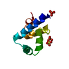
| ||||||||
|---|---|---|---|---|---|---|---|---|---|
| 1 | 
| ||||||||
| Unit cell |
| ||||||||
| Details | The second part of the biological assembly is generated by the two fold axis: -x+1, -y, z+3/2 |
-Components
| #1: Protein | / BldD Mass: 7883.973 Da / Num. of mol.: 1 / Fragment: DNA-binding domain Source method: isolated from a genetically manipulated source Source: (gene. exp.) Streptomyces coelicolor (bacteria) / Strain: A3(2) / Plasmid: pET15b / Species (production host): Escherichia coli / Production host: Escherichia coli BL21 (bacteria) / Strain (production host): BL21 / References: GenBank: 21219990, UniProt: Q7AKQ8*PLUS | ||
|---|---|---|---|
| #2: Chemical | Sulfate  Mass: 96.063 Da / Num. of mol.: 3 / Source method: obtained synthetically / Formula: SO4 Mass: 96.063 Da / Num. of mol.: 3 / Source method: obtained synthetically / Formula: SO4#3: Water | ChemComp-HOH / | Water Mass: 18.015 Da / Num. of mol.: 147 / Source method: isolated from a natural source / Formula: H2O Mass: 18.015 Da / Num. of mol.: 147 / Source method: isolated from a natural source / Formula: H2O |
-Experimental details
-Experiment
| Experiment | Method: X-RAY DIFFRACTION / Number of used crystals: 1 |
|---|
- Sample preparation
| Crystal | Density Matthews: 2.53 Å3/Da / Density % sol: 51.29 % |
|---|---|
| Crystal grow | Temperature: 296 K / Method: vapor diffusion, hanging drop / pH: 4.6 Details: 25% PEG 4000, 0.1M sodium acetate (pH 4.6), 0.2M ammonium sulfate, VAPOR DIFFUSION, HANGING DROP, temperature 296K |
-Data collection
| Diffraction | Mean temperature: 100 K | ||||||||||||
|---|---|---|---|---|---|---|---|---|---|---|---|---|---|
| Diffraction source | Source: SYNCHROTRON / Site: PAL/PLS  / Beamline: 6B / Wavelength: 0.97932, 0.97942, 0.98722 / Beamline: 6B / Wavelength: 0.97932, 0.97942, 0.98722 | ||||||||||||
| Detector | Type: MAC Science DIP-2030B / Detector: IMAGE PLATE / Date: Dec 5, 2003 | ||||||||||||
| Radiation | Monochromator: Si / Protocol: MAD / Monochromatic (M) / Laue (L): M / Scattering type: x-ray | ||||||||||||
| Radiation wavelength |
| ||||||||||||
| Reflection | Resolution: 1.8→20 Å / Num. all: 7441 / Num. obs: 7031 / % possible obs: 94.5 % / Observed criterion σ(F): 0 / Observed criterion σ(I): 0 / Biso Wilson estimate: 14.7 Å2 | ||||||||||||
| Reflection shell | Resolution: 1.8→1.86 Å / % possible all: 94.9 |
- Processing
| Software |
| |||||||||||||||||||||||||
|---|---|---|---|---|---|---|---|---|---|---|---|---|---|---|---|---|---|---|---|---|---|---|---|---|---|---|
| Refinement | Method to determine structure: MAD / Resolution: 1.81→19.57 Å / Rfactor Rfree error: 0.012 / Data cutoff high absF: 1438212.22 / Data cutoff low absF: 0 / Isotropic thermal model: RESTRAINED / Cross valid method: THROUGHOUT / σ(F): 0 / Stereochemistry target values: Engh & Huber
| |||||||||||||||||||||||||
| Solvent computation | Solvent model: FLAT MODEL / Bsol: 54.3987 Å2 / ksol: 0.351951 e/Å3 | |||||||||||||||||||||||||
| Displacement parameters | Biso mean: 18.3 Å2
| |||||||||||||||||||||||||
| Refine analyze |
| |||||||||||||||||||||||||
| Refinement step | Cycle: LAST / Resolution: 1.81→19.57 Å
| |||||||||||||||||||||||||
| Refine LS restraints |
| |||||||||||||||||||||||||
| LS refinement shell | Resolution: 1.8→1.91 Å / Total num. of bins used: 6
| |||||||||||||||||||||||||
| Xplor file |
|
