 Movie
Movie Controller


+ Open data

- Basic information
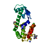
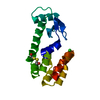
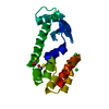
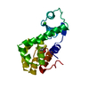
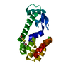
| Entry | Database: PDB / ID: 2zug | ||||||
|---|---|---|---|---|---|---|---|
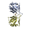
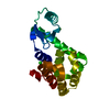
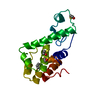
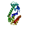
| Title | Crystal structure of WSSV ICP11 | ||||||
 Components Components | ORF115 (WSSV285) (Wsv230) | ||||||
 Keywords Keywords |  VIRAL PROTEIN / DNA mimic protein / dimer / White spot syndrome virus VIRAL PROTEIN / DNA mimic protein / dimer / White spot syndrome virus | ||||||
| Function / homology | VP9 protein domain / White spot syndrome virus (WSSV), Orf116/126, N-terminal / VP9, N-terminal domain superfamily / VP9 protein / Alpha-Beta Plaits / 2-Layer Sandwich / Alpha Beta / ICP11/P9 Function and homology information Function and homology information | ||||||
| Biological species |  Shrimp white spot syndrome virus Shrimp white spot syndrome virus | ||||||
| Method | X-RAY DIFFRACTION / SYNCHROTRON / MAD / Resolution: 2.72 Å | ||||||
 Authors Authors | Wang, A.H.-J. / Wang, H.-C. / Ko, T.-P. / Lo, C.-F. | ||||||
 Citation Citation | Journal: Proc.Natl.Acad.Sci.USA / Year: 2008 Title: White spot syndrome virus protein ICP11: A histone-binding DNA mimic that disrupts nucleosome assembly Authors: Wang, H.-C. / Wang, H.-C. / Ko, T.-P. / Lee, Y.-M. / Leu, J.-H. / Ho, C.-H. / Huang, W.-P. / Lo, C.-F. / Wang, A.H.-J. | ||||||
| History |
|
- Structure visualization
| Structure viewer | Molecule: MolmilJmol/JSmol |
|---|
- Downloads & links
-Download
| PDBx/mmCIF format | 2zug.cif.gz | 39.3 KB | Display | PDBx/mmCIF format |
|---|---|---|---|---|
| PDB format | pdb2zug.ent.gz | 31.7 KB | Display | PDB format |
| PDBx/mmJSON format | 2zug.json.gz | Tree view | PDBx/mmJSON format | |
| Others |  Other downloads Other downloads |
-Validation report
| Arichive directory | https://data.pdbj.org/pub/pdb/validation_reports/zu/2zugftp://data.pdbj.org/pub/pdb/validation_reports/zu/2zug | HTTPS FTP |
|---|
-Related structure data
| Related structure data | |
|---|---|
| Similar structure data |

-Links
 PDBj
PDBj- Assembly
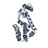
| Deposited unit | 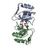
| ||||||||||||
|---|---|---|---|---|---|---|---|---|---|---|---|---|---|
| 1 |
| ||||||||||||
| Unit cell |
| ||||||||||||
| Components on special symmetry positions |
|
-Components
| #1: Protein | Mass: 10477.116 Da / Num. of mol.: 2 Source method: isolated from a genetically manipulated source Source: (gene. exp.) Shrimp white spot syndrome virus / Strain: Taiwan isolate / Gene: white spot syndrome virus / Plasmid: pET21b / Production host:  Escherichia coli (E. coli) / Strain (production host): BL21 (DE3) / References: UniProt: Q91LD0 Escherichia coli (E. coli) / Strain (production host): BL21 (DE3) / References: UniProt: Q91LD0#2: Water | ChemComp-HOH / | Water Mass: 18.015 Da / Num. of mol.: 39 / Source method: isolated from a natural source / Formula: H2O Mass: 18.015 Da / Num. of mol.: 39 / Source method: isolated from a natural source / Formula: H2O |
|---|
-Experimental details
-Experiment
| Experiment | Method: X-RAY DIFFRACTION / Number of used crystals: 2 |
|---|
- Sample preparation
| Crystal | Density Matthews: 4.92 Å3/Da / Density % sol: 75.01 % |
|---|---|
| Crystal grow | Temperature: 298 K / Method: vapor diffusion, sitting drop / pH: 6.5 Details: 0.2M sodium acetate trihydrate, 2.2M ammonium sulfate, pH 6.5, VAPOR DIFFUSION, SITTING DROP, temperature 298K |
-Data collection
| Diffraction |
| ||||||||||||||||||
|---|---|---|---|---|---|---|---|---|---|---|---|---|---|---|---|---|---|---|---|
| Diffraction source |
| ||||||||||||||||||
| Detector |
| ||||||||||||||||||
| Radiation |
| ||||||||||||||||||
| Radiation wavelength |
| ||||||||||||||||||
| Reflection | Resolution: 2.72→30 Å / Num. all: 11756 / Num. obs: 11437 / % possible obs: 96.8 % / Observed criterion σ(F): 0 / Observed criterion σ(I): 1 / Redundancy: 9.4 % / Rmerge(I) obs: 0.062 / Net I/σ(I): 30.3 | ||||||||||||||||||
| Reflection shell | Resolution: 2.72→2.82 Å / Redundancy: 10.1 % / Rmerge(I) obs: 0.644 / Mean I/σ(I) obs: 3.2 / Num. unique all: 1158 / % possible all: 88.2 |

- Processing
| Software |
| |||||||||||||||||||||||||
|---|---|---|---|---|---|---|---|---|---|---|---|---|---|---|---|---|---|---|---|---|---|---|---|---|---|---|
| Refinement | Method to determine structure: MAD / Resolution: 2.72→29.25 Å / Isotropic thermal model: Isotropic / Cross valid method: THROUGHOUT / σ(F): 0 / σ(I): 0 / Stereochemistry target values: Engh & Huber
| |||||||||||||||||||||||||
| Displacement parameters | Biso mean: 66.8 Å2 | |||||||||||||||||||||||||
| Refine analyze |
| |||||||||||||||||||||||||
| Refinement step | Cycle: LAST / Resolution: 2.72→29.25 Å
| |||||||||||||||||||||||||
| Refine LS restraints |
| |||||||||||||||||||||||||
| LS refinement shell | Resolution: 2.72→2.82 Å / Rfactor Rfree error: 0.035
|
