Method to determine structure: MAD / Resolution: 2.28→26.7 Å / Cor.coef. Fo:Fc: 0.934 / Cor.coef. Fo:Fc free: 0.913 / SU B: 13.938 / SU ML: 0.175 / TLS residual ADP flag: LIKELY RESIDUAL / Cross valid method: THROUGHOUT / ESU R: 0.318 / ESU R Free: 0.25 Stereochemistry target values: MAXIMUM LIKELIHOOD WITH PHASES Details: 1. HYDROGENS HAVE BEEN ADDED IN THE RIDING POSITIONS 2. A MET-INHIBITION PROTOCOL WAS USED FOR SELENOMETHIONINE INCORPORATION DURING PROTEIN EXPRESSION. THE OCCUPANCY OF THE SE ATOMS IN THE ...Details: 1. HYDROGENS HAVE BEEN ADDED IN THE RIDING POSITIONS 2. A MET-INHIBITION PROTOCOL WAS USED FOR SELENOMETHIONINE INCORPORATION DURING PROTEIN EXPRESSION. THE OCCUPANCY OF THE SE ATOMS IN THE MSE RESIDUES WAS REDUCED TO 0.7 TO ACCOUNT FOR THE REDUCED SCATTERING POWER DUE TO PARTIAL S-MET INCORPORATION. 3. THE STRUCTURE WAS NOT MODELED BETWEEN 162-171 BECAUSE THE ELECTRON DENSITY IN THIS REGION IS DISORDERED. 4. THIS PROTEIN IS REDUCTIVELY METHYLATED. HOWEVER, DENSITY FOR MOST OF THE METHYL GROUPS ARE NOT OBSERVED, MAYBE DUE TO FLEXIBILITY OR LIMITED RESOLUTION.
Rfactor
Num. reflection
% reflection
Selection details
Rfree
0.27
375
4.6 %
RANDOM
Rwork
0.216
-
-
-
all
0.219
-
-
-
obs
-
7717
93.03 %
-
Solvent computation
Ion probe radii: 0.8 Å / Shrinkage radii: 0.8 Å / VDW probe radii: 1.2 Å / Solvent model: BABINET MODEL WITH MASK
Displacement parameters
Biso mean: 40.488 Å2
Baniso -1
Baniso -2
Baniso -3
1-
-1.66 Å2
0 Å2
0 Å2
2-
-
1.78 Å2
0 Å2
3-
-
-
-0.11 Å2
Refinement step
Cycle: LAST / Resolution: 2.28→26.7 Å
Protein
Nucleic acid
Ligand
Solvent
Total
Num. atoms
1106
0
1
62
1169
Refine LS restraints
Refine-ID
Type
Dev ideal
Dev ideal target
Number
X-RAY DIFFRACTION
r_bond_refined_d
0.016
0.022
1119
X-RAY DIFFRACTION
r_bond_other_d
0.001
0.02
1023
X-RAY DIFFRACTION
r_angle_refined_deg
1.362
1.968
1505
X-RAY DIFFRACTION
r_angle_other_deg
0.835
3
2384
X-RAY DIFFRACTION
r_dihedral_angle_1_deg
5.25
5
139
X-RAY DIFFRACTION
r_dihedral_angle_2_deg
31.773
25.263
57
X-RAY DIFFRACTION
r_dihedral_angle_3_deg
15.049
15
208
X-RAY DIFFRACTION
r_dihedral_angle_4_deg
24.674
15
8
X-RAY DIFFRACTION
r_chiral_restr
0.076
0.2
168
X-RAY DIFFRACTION
r_gen_planes_refined
0.005
0.02
1246
X-RAY DIFFRACTION
r_gen_planes_other
0.001
0.02
210
X-RAY DIFFRACTION
r_nbd_refined
0.218
0.2
259
X-RAY DIFFRACTION
r_nbd_other
0.168
0.2
927
X-RAY DIFFRACTION
r_nbtor_refined
0.182
0.2
540
X-RAY DIFFRACTION
r_nbtor_other
0.085
0.2
656
X-RAY DIFFRACTION
r_xyhbond_nbd_refined
0.199
0.2
41
X-RAY DIFFRACTION
r_symmetry_vdw_refined
0.181
0.2
9
X-RAY DIFFRACTION
r_symmetry_vdw_other
0.386
0.2
18
X-RAY DIFFRACTION
r_symmetry_hbond_refined
0.133
0.2
6
X-RAY DIFFRACTION
r_mcbond_it
2.243
3
735
X-RAY DIFFRACTION
r_mcbond_other
0.562
3
284
X-RAY DIFFRACTION
r_mcangle_it
3.354
5
1116
X-RAY DIFFRACTION
r_scbond_it
5.965
8
449
X-RAY DIFFRACTION
r_scangle_it
8.617
11
389
LS refinement shell
Resolution: 2.28→2.339 Å / Total num. of bins used: 20
Rfactor
Num. reflection
% reflection
Rfree
0.391
22
-
Rwork
0.24
568
-
obs
-
-
93.21 %
Refinement TLS params.
Method: refined / Origin x: 14.1683 Å / Origin y: 28.8656 Å / Origin z: 53.6444 Å
11
12
13
21
22
23
31
32
33
T
-0.1623 Å2
-0.0042 Å2
-0.0423 Å2
-
-0.2821 Å2
0.0024 Å2
-
-
-0.124 Å2
L
3.4018 °2
0.1682 °2
-2.1498 °2
-
1.0196 °2
-0.8106 °2
-
-
6.6168 °2
S
0.0338 Å °
-0.0457 Å °
0.2954 Å °
0.0509 Å °
0.0258 Å °
-0.0354 Å °
-0.3101 Å °
0.1317 Å °
-0.0596 Å °
+
About Yorodumi
-
News
-
Feb 9, 2022. New format data for meta-information of EMDB entries
New format data for meta-information of EMDB entries
Version 3 of the EMDB header file is now the official format.
The previous official version 1.9 will be removed from the archive.
In the structure databanks used in Yorodumi, some data are registered as the other names, "COVID-19 virus" and "2019-nCoV". Here are the details of the virus and the list of structure data.
Jan 31, 2019. EMDB accession codes are about to change! (news from PDBe EMDB page)
EMDB accession codes are about to change! (news from PDBe EMDB page)
The allocation of 4 digits for EMDB accession codes will soon come to an end. Whilst these codes will remain in use, new EMDB accession codes will include an additional digit and will expand incrementally as the available range of codes is exhausted. The current 4-digit format prefixed with “EMD-” (i.e. EMD-XXXX) will advance to a 5-digit format (i.e. EMD-XXXXX), and so on. It is currently estimated that the 4-digit codes will be depleted around Spring 2019, at which point the 5-digit format will come into force.
The EM Navigator/Yorodumi systems omit the EMD- prefix.
Related info.:Q: What is EMD? / ID/Accession-code notation in Yorodumi/EM Navigator
Yorodumi is a browser for structure data from EMDB, PDB, SASBDB, etc.
This page is also the successor to EM Navigator detail page, and also detail information page/front-end page for Omokage search.
The word "yorodu" (or yorozu) is an old Japanese word meaning "ten thousand". "mi" (miru) is to see.
Related info.:EMDB / PDB / SASBDB / Comparison of 3 databanks / Yorodumi Search / Aug 31, 2016. New EM Navigator & Yorodumi / Yorodumi Papers / Jmol/JSmol / Function and homology information / Changes in new EM Navigator and Yorodumi
 Movie
Movie Controller

 Yorodumi
 Open data
 Basic information
 Components
 Keywords
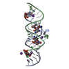
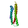
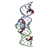
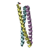
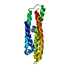
 MEMBRANE PROTEIN / Bromodomain-like fold /
  Function and homology information

 Authors
 Citation
 Structure visualization
 Downloads & links
 Other downloads

 PDBj
PDBj Assembly
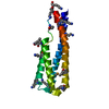

 Mass: 35.453 Da / Num. of mol.: 1 / Source method: obtained synthetically / Formula: Cl
 Mass: 18.015 Da / Num. of mol.: 62 / Source method: isolated from a natural source / Formula: H2O
 Sample preparation
 / Beamline: 8.2.2 / Wavelength: 1.0163, 0.9797, 0.9796
 Processing