 Movie
Movie Controller

[English] 日本語
 Yorodumi
- PDB-3dvv: Crystal structure of HIV-1 subtype F DIS extended duplex RNA boun... -
+ Open data

- Basic information
| Entry | Database: PDB / ID: 3dvv | ||||||
|---|---|---|---|---|---|---|---|
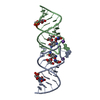
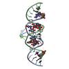
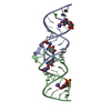
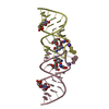
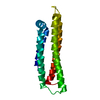
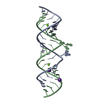
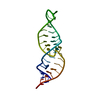
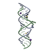
| Title | Crystal structure of HIV-1 subtype F DIS extended duplex RNA bound to ribostamycin (U267OMe) | ||||||
 Components Components | HIV-1 genomic RNA | ||||||
 Keywords Keywords |  RNA / HIV-1 / antibiotic / aminoglycoside RNA / HIV-1 / antibiotic / aminoglycoside | ||||||
| Function / homology | : / RIBOSTAMYCIN / RNA / RNA (> 10) Function and homology information Function and homology information | ||||||
| Method | X-RAY DIFFRACTION / SYNCHROTRON / MOLECULAR REPLACEMENT / Resolution: 2 Å | ||||||
 Authors Authors | Ennifar, E. / Dumas, P. / Olieric, V. | ||||||
 Citation Citation | Journal: To be Published Title: A fast strategy for Selenium derivatization and phasing of RNA structures Authors: Olieric, V. / Rieder, U. / Lang, K. / Serganov, A. / Schulze-Brise, C. / Micura, R. / Dumas, P. / Ennifar, E. | ||||||
| History |
|
- Structure visualization
| Structure viewer | Molecule: MolmilJmol/JSmol |
|---|
- Downloads & links
-Download
| PDBx/mmCIF format | 3dvv.cif.gz | 40.3 KB | Display | PDBx/mmCIF format |
|---|---|---|---|---|
| PDB format | pdb3dvv.ent.gz | 27.3 KB | Display | PDB format |
| PDBx/mmJSON format | 3dvv.json.gz | Tree view | PDBx/mmJSON format | |
| Others |  Other downloads Other downloads |
-Validation report
| Arichive directory | https://data.pdbj.org/pub/pdb/validation_reports/dv/3dvvftp://data.pdbj.org/pub/pdb/validation_reports/dv/3dvv | HTTPS FTP |
|---|
-Related structure data
| Related structure data |  3c3zS S: Starting model for refinement |
|---|---|
| Similar structure data |


-Links
 PDBj
PDBj


- Assembly
| Deposited unit | 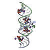
| ||||||||
|---|---|---|---|---|---|---|---|---|---|
| 1 |
| ||||||||
| Unit cell |
|
-Components
| #1: RNA chain | Mass: 7400.499 Da / Num. of mol.: 2 / Source method: obtained synthetically #2: Chemical | ChemComp-K /   Mass: 39.098 Da / Num. of mol.: 4 / Source method: obtained synthetically / Formula: K Mass: 39.098 Da / Num. of mol.: 4 / Source method: obtained synthetically / Formula: K#3: Chemical | ChemComp-CL / | Chloride  Mass: 35.453 Da / Num. of mol.: 1 / Source method: obtained synthetically / Formula: Cl Mass: 35.453 Da / Num. of mol.: 1 / Source method: obtained synthetically / Formula: Cl#4: Chemical | Ribostamycin  Mass: 454.473 Da / Num. of mol.: 2 / Source method: obtained synthetically / Formula: C17H34N4O10 / Comment: antibiotic*YM Mass: 454.473 Da / Num. of mol.: 2 / Source method: obtained synthetically / Formula: C17H34N4O10 / Comment: antibiotic*YM#5: Water | ChemComp-HOH / | Water Mass: 18.015 Da / Num. of mol.: 44 / Source method: isolated from a natural source / Formula: H2O Mass: 18.015 Da / Num. of mol.: 44 / Source method: isolated from a natural source / Formula: H2O |
|---|
-Experimental details
-Experiment
| Experiment | Method: X-RAY DIFFRACTION / Number of used crystals: 1 |
|---|
- Sample preparation
| Crystal | Density Matthews: 2.52 Å3/Da / Density % sol: 51.17 % | ||||||||||||||||||||||||||||||||||||
|---|---|---|---|---|---|---|---|---|---|---|---|---|---|---|---|---|---|---|---|---|---|---|---|---|---|---|---|---|---|---|---|---|---|---|---|---|---|
| Crystal grow | Temperature: 310 K / Method: vapor diffusion, sitting drop / pH: 6.2 Details: MPD, cacodylate, MgCl2, KCl, pH 6.2, VAPOR DIFFUSION, SITTING DROP, temperature 310K | ||||||||||||||||||||||||||||||||||||
| Components of the solutions |
|
-Data collection
| Diffraction | Mean temperature: 90 K |
|---|---|
| Diffraction source | Source: SYNCHROTRON / Site: ESRF  / Beamline: ID29 / Wavelength: 0.92 Å / Beamline: ID29 / Wavelength: 0.92 Å |
| Detector | Type: ADSC QUANTUM 315 / Detector: CCD / Date: Jul 13, 2007 |
| Radiation | Protocol: SINGLE WAVELENGTH / Monochromatic (M) / Laue (L): M / Scattering type: x-ray |
| Radiation wavelength | Wavelength: 0.92 Å / Relative weight: 1 |
| Reflection | Resolution: 2→30 Å / Num. all: 10096 / Num. obs: 9829 / % possible obs: 95.6 % / Observed criterion σ(F): 0 / Observed criterion σ(I): 0 / Redundancy: 8.2 % / Biso Wilson estimate: 47.6 Å2 / Rsym value: 0.058 / Net I/σ(I): 29 |
| Reflection shell | Resolution: 2→2.07 Å / Redundancy: 8 % / Mean I/σ(I) obs: 9.8 / Num. unique all: 971 / Rsym value: 0.309 / % possible all: 91.3 |
- Processing
| Software |
| ||||||||||||||||||||||||||||||||||||||||||||||||||||||||||||
|---|---|---|---|---|---|---|---|---|---|---|---|---|---|---|---|---|---|---|---|---|---|---|---|---|---|---|---|---|---|---|---|---|---|---|---|---|---|---|---|---|---|---|---|---|---|---|---|---|---|---|---|---|---|---|---|---|---|---|---|---|---|
| Refinement | Method to determine structure: MOLECULAR REPLACEMENT Starting model: PDB ID 3C3Z Resolution: 2→18.59 Å / Rfactor Rfree error: 0.01 / Data cutoff high absF: 741857.93 / Data cutoff low absF: 0 / Isotropic thermal model: RESTRAINED / Cross valid method: THROUGHOUT / σ(F): 0
| ||||||||||||||||||||||||||||||||||||||||||||||||||||||||||||
| Solvent computation | Solvent model: FLAT MODEL / Bsol: 36.979 Å2 / ksol: 0.358534 e/Å3 | ||||||||||||||||||||||||||||||||||||||||||||||||||||||||||||
| Displacement parameters | Biso mean: 45.1 Å2
| ||||||||||||||||||||||||||||||||||||||||||||||||||||||||||||
| Refine analyze |
| ||||||||||||||||||||||||||||||||||||||||||||||||||||||||||||
| Refinement step | Cycle: LAST / Resolution: 2→18.59 Å
| ||||||||||||||||||||||||||||||||||||||||||||||||||||||||||||
| Refine LS restraints |
| ||||||||||||||||||||||||||||||||||||||||||||||||||||||||||||
| LS refinement shell | Resolution: 2→2.13 Å / Rfactor Rfree error: 0.038 / Total num. of bins used: 6
| ||||||||||||||||||||||||||||||||||||||||||||||||||||||||||||
| Xplor file |
|
