 ムービー
ムービー コントローラー


+ データを開く

- 基本情報
基本情報
| 登録情報 | データベース: PDB / ID: 2elb | ||||||
|---|---|---|---|---|---|---|---|
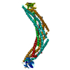
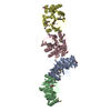
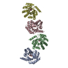
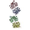
| タイトル | Crystal Structure of the BAR-PH domain of human APPL1 | ||||||
 要素 要素 | Adapter protein containing PH domain, PTB domain and leucine zipper motif 1 | ||||||
 キーワード キーワード | PROTEIN BINDING (タンパク質) / APPL / BAR domain / PH domain | ||||||
| 機能・相同性 |  機能・相同性情報 機能・相同性情報negative regulation of Fc-gamma receptor signaling pathway involved in phagocytosis / positive regulation of macropinocytosis / adiponectin-activated signaling pathway / マクロピノソーム / regulation of fibroblast migration / regulation of glucose import / maintenance of synapse structure / protein kinase B binding / signaling / positive regulation of melanin biosynthetic process ...negative regulation of Fc-gamma receptor signaling pathway involved in phagocytosis / positive regulation of macropinocytosis / adiponectin-activated signaling pathway / マクロピノソーム / regulation of fibroblast migration / regulation of glucose import / maintenance of synapse structure / protein kinase B binding / signaling / positive regulation of melanin biosynthetic process / regulation of toll-like receptor 4 signaling pathway / vesicle membrane / early phagosome / Caspase activation via Dependence Receptors in the absence of ligand / positive regulation of cytokine production involved in inflammatory response / cellular response to hepatocyte growth factor stimulus / intracellular vesicle / regulation of innate immune response / beta-tubulin binding / phosphatidylserine binding / regulation of G1/S transition of mitotic cell cycle / regulation of protein localization to plasma membrane / ruffle / phosphatidylinositol binding / transforming growth factor beta receptor signaling pathway / positive regulation of glucose import / protein import into nucleus / presynapse / insulin receptor signaling pathway / early endosome membrane / cytoplasmic vesicle / postsynapse / エンドソーム / endosome membrane / エンドソーム / 細胞周期 / glutamatergic synapse / protein-containing complex binding / シグナル伝達 / protein homodimerization activity / extracellular exosome / 生体膜 / identical protein binding / 細胞核 / 細胞膜 / 細胞質基質 / 細胞質類似検索 - 分子機能 | ||||||
| 生物種 |  Homo sapiens (ヒト) Homo sapiens (ヒト) | ||||||
| 手法 | X線回折 / シンクロトロン / 単波長異常分散 / 解像度: 2.6 Å | ||||||
 データ登録者 データ登録者 | Li, J. / Mao, X. / Dong, L.Q. / Liu, F. / Tong, L. | ||||||
 引用 引用 | ジャーナル: Structure / 年: 2007 タイトル: Crystal Structures of the BAR-PH and PTB Domains of Human APPL1 著者: Li, J. / Mao, X. / Dong, L.Q. / Liu, F. / Tong, L. | ||||||
| 履歴 |
|
- 構造の表示
構造の表示
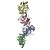
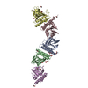
| 構造ビューア | 分子: MolmilJmol/JSmol |
|---|
- ダウンロードとリンク
-ダウンロード
| PDBx/mmCIF形式 | 2elb.cif.gz | 80 KB | 表示 | PDBx/mmCIF形式 |
|---|---|---|---|---|
| PDB形式 | pdb2elb.ent.gz | 64.2 KB | 表示 | PDB形式 |
| PDBx/mmJSON形式 | 2elb.json.gz | ツリー表示 | PDBx/mmJSON形式 | |
| その他 |  その他のダウンロード その他のダウンロード |
-検証レポート
| アーカイブディレクトリ | https://data.pdbj.org/pub/pdb/validation_reports/el/2elbftp://data.pdbj.org/pub/pdb/validation_reports/el/2elb | HTTPS FTP |
|---|
-関連構造データ


-リンク
 PDBj
PDBj


- 集合体
集合体
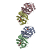
| 登録構造単位 | 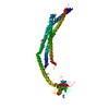
| ||||||||
|---|---|---|---|---|---|---|---|---|---|
| 1 | 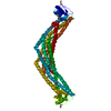
| ||||||||
| 単位格子 |
| ||||||||
| 詳細 | The biological assembly is a dimer. |
-要素
| #1: タンパク質 | / APPL1 / Dip13 alpha / DCC-interacting protein 13 alpha 分子量: 45987.117 Da / 分子数: 1 / 断片: The BAR-PH domain / 由来タイプ: 組換発現 / 由来: (組換発現) Homo sapiens (ヒト) / プラスミド: pET28a / 生物種 (発現宿主): Escherichia coli / 発現宿主:  Escherichia coli BL21(DE3) (大腸菌) / 株 (発現宿主): BL21(DE3) / 参照: UniProt: Q9UKG1 Escherichia coli BL21(DE3) (大腸菌) / 株 (発現宿主): BL21(DE3) / 参照: UniProt: Q9UKG1 |
|---|
-実験情報
-実験
| 実験 | 手法: X線回折 / 使用した結晶の数: 1 |
|---|
- 試料調製
試料調製
| 結晶 | マシュー密度: 2.11 Å3/Da / 溶媒含有率: 41.7 % |
|---|---|
| 結晶化 | 温度: 293 K / 手法: 蒸気拡散法, シッティングドロップ法 / pH: 7.5 詳細: 0.1M HEPES (pH 7.5), 22% (w/v) polyacrylic acid 5100, 20mM magnesium chloride, VAPOR DIFFUSION, SITTING DROP, temperature 293K |
-データ収集
| 回折 | 平均測定温度: 100 K |
|---|---|
| 放射光源 | 由来: シンクロトロン / サイト: NSLS  / ビームライン: X4A / 波長: 0.97934 Å / ビームライン: X4A / 波長: 0.97934 Å |
| 検出器 | タイプ: ADSC QUANTUM 4 / 検出器: CCD / 詳細: mirrors |
| 放射 | プロトコル: SINGLE WAVELENGTH / 単色(M)・ラウエ(L): M / 散乱光タイプ: x-ray |
| 放射波長 | 波長: 0.97934 Å / 相対比: 1 |
| 反射 | 解像度: 2.6→30 Å / Num. obs: 22741 / % possible obs: 98.3 % / Observed criterion σ(F): 2 / Observed criterion σ(I): 1 / 冗長度: 6.4 % / Biso Wilson estimate: 29.1 Å2 / Rmerge(I) obs: 0.067 / Net I/σ(I): 27.5853 |
| 反射 シェル | 解像度: 2.6→2.69 Å / 冗長度: 6 % / Rmerge(I) obs: 0.339 / Mean I/σ(I) obs: 5.94 / Num. unique all: 2267 / % possible all: 98.4 |
- 解析
解析
| ソフトウェア |
| ||||||||||||||||||||||||||||||||||||||||||||||||||||||||||||||||||||||||||||||||
|---|---|---|---|---|---|---|---|---|---|---|---|---|---|---|---|---|---|---|---|---|---|---|---|---|---|---|---|---|---|---|---|---|---|---|---|---|---|---|---|---|---|---|---|---|---|---|---|---|---|---|---|---|---|---|---|---|---|---|---|---|---|---|---|---|---|---|---|---|---|---|---|---|---|---|---|---|---|---|---|---|---|
| 精密化 | 構造決定の手法: 単波長異常分散 / 解像度: 2.6→29.84 Å / Rfactor Rfree error: 0.009 / Data cutoff high absF: 594181.77 / Data cutoff low absF: 0 / Isotropic thermal model: RESTRAINED / 交差検証法: THROUGHOUT / σ(F): 1.5
| ||||||||||||||||||||||||||||||||||||||||||||||||||||||||||||||||||||||||||||||||
| 溶媒の処理 | 溶媒モデル: FLAT MODEL / Bsol: 22.6318 Å2 / ksol: 0.304871 e/Å3 | ||||||||||||||||||||||||||||||||||||||||||||||||||||||||||||||||||||||||||||||||
| 原子変位パラメータ | Biso mean: 44.9 Å2
| ||||||||||||||||||||||||||||||||||||||||||||||||||||||||||||||||||||||||||||||||
| Refine analyze |
| ||||||||||||||||||||||||||||||||||||||||||||||||||||||||||||||||||||||||||||||||
| 精密化ステップ | サイクル: LAST / 解像度: 2.6→29.84 Å
| ||||||||||||||||||||||||||||||||||||||||||||||||||||||||||||||||||||||||||||||||
| 拘束条件 |
| ||||||||||||||||||||||||||||||||||||||||||||||||||||||||||||||||||||||||||||||||
| LS精密化 シェル | 解像度: 2.6→2.69 Å / Rfactor Rfree error: 0.032 / Total num. of bins used: 10
| ||||||||||||||||||||||||||||||||||||||||||||||||||||||||||||||||||||||||||||||||
| Xplor file |
|
