 Movie
Movie Controller


+ Open data

- Basic information
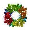
| Entry | Database: PDB / ID: 2wl3 | ||||||
|---|---|---|---|---|---|---|---|
| Title | crystal structure of catechol 2,3-dioxygenase | ||||||
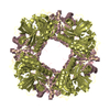
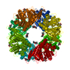
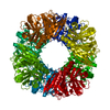
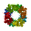
 Components Components | CATECHOL 2,3-DIOXYGENASE | ||||||
 Keywords Keywords | OXIDOREDUCTASE / AROMATIC HYDROCARBONS CATABOLISM / AKBC | ||||||
| Function / homology |  Function and homology information Function and homology information: / dioxygenase activity / xenobiotic catabolic process / ferrous iron bindingSimilarity search - Function | ||||||
| Biological species |  RHODOCOCCUS SP. DK17 (bacteria) RHODOCOCCUS SP. DK17 (bacteria) | ||||||
| Method | X-RAY DIFFRACTION / SYNCHROTRON / SAD / Resolution: 2.2 Å | ||||||
 Authors Authors | Cho, H.J. / Kim, K.J. / Kang, B.S. | ||||||
 Citation Citation | Journal: J.Biol.Chem. / Year: 2010 Title: Substrate-Binding Mechanism of a Type I Extradiol Dioxygenase. Authors: Cho, H.J. / Kim, K. / Sohn, S.Y. / Cho, H.Y. / Kim, K.J. / Kim, M.H. / Kim, D. / Kim, E. / Kang, B.S. | ||||||
| History |
| ||||||
| Remark 700 | SHEET THE SHEET STRUCTURE OF THIS MOLECULE IS BIFURCATED. IN ORDER TO REPRESENT THIS FEATURE IN ... SHEET THE SHEET STRUCTURE OF THIS MOLECULE IS BIFURCATED. IN ORDER TO REPRESENT THIS FEATURE IN THE SHEET RECORDS BELOW, TWO SHEETS ARE DEFINED. |
- Structure visualization
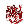
| Structure viewer | Molecule: MolmilJmol/JSmol |
|---|
- Downloads & links
-Download
| PDBx/mmCIF format | 2wl3.cif.gz | 441.4 KB | Display | PDBx/mmCIF format |
|---|---|---|---|---|
| PDB format | pdb2wl3.ent.gz | 380.6 KB | Display | PDB format |
| PDBx/mmJSON format | 2wl3.json.gz | Tree view | PDBx/mmJSON format | |
| Others |  Other downloads Other downloads |
-Validation report
| Arichive directory | https://data.pdbj.org/pub/pdb/validation_reports/wl/2wl3ftp://data.pdbj.org/pub/pdb/validation_reports/wl/2wl3 | HTTPS FTP |
|---|
-Related structure data


-Links
 PDBj
PDBj- Assembly
| Deposited unit | 
| ||||||||
|---|---|---|---|---|---|---|---|---|---|
| 1 | 
| ||||||||
| 2 | 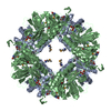
| ||||||||
| Unit cell |
|
-Components
| #1: Protein | Mass: 34443.336 Da / Num. of mol.: 4 Source method: isolated from a genetically manipulated source Source: (gene. exp.) RHODOCOCCUS SP. DK17 (bacteria) / Production host: ESCHERICHIA COLI (E. coli) / Strain (production host): B834 / References: UniProt: Q6REQ5#2: Chemical | ChemComp-FE / Iron  Mass: 55.845 Da / Num. of mol.: 4 / Source method: obtained synthetically / Formula: Fe Mass: 55.845 Da / Num. of mol.: 4 / Source method: obtained synthetically / Formula: Fe#3: Chemical | ChemComp-CA / |   Mass: 40.078 Da / Num. of mol.: 1 / Source method: obtained synthetically / Formula: Ca Mass: 40.078 Da / Num. of mol.: 1 / Source method: obtained synthetically / Formula: Ca#4: Chemical | ChemComp-GOL / Glycerol  Mass: 92.094 Da / Num. of mol.: 5 / Source method: obtained synthetically / Formula: C3H8O3 Mass: 92.094 Da / Num. of mol.: 5 / Source method: obtained synthetically / Formula: C3H8O3#5: Water | ChemComp-HOH / | Water Mass: 18.015 Da / Num. of mol.: 251 / Source method: isolated from a natural source / Formula: H2O Mass: 18.015 Da / Num. of mol.: 251 / Source method: isolated from a natural source / Formula: H2ONonpolymer details | SELENOMETH | Sequence details | ABOUT 20 RESIDUES AT C-TERMINUS ARE DELETED | |
|---|
-Experimental details
-Experiment
| Experiment | Method: X-RAY DIFFRACTION |
|---|
- Sample preparation
| Crystal | Density Matthews: 2.74 Å3/Da / Density % sol: 55 % / Description: NONE |
|---|---|
| Crystal grow | Details: 30% PEG 400, 0.1M HEPES PH7.5, 0.2M CALCIUM CHLORIDE |
-Data collection
| Diffraction | Mean temperature: 294 K | |||||||||||||||
|---|---|---|---|---|---|---|---|---|---|---|---|---|---|---|---|---|
| Diffraction source | Source: SYNCHROTRON / Site: PAL/PLS  / Beamline: 4A / Wavelength: 0.9796 / Beamline: 4A / Wavelength: 0.9796 | |||||||||||||||
| Detector | Type: ADSC CCD / Detector: CCD / Date: Jul 30, 2005 | |||||||||||||||
| Radiation | Protocol: SINGLE WAVELENGTH / Monochromatic (M) / Laue (L): M / Scattering type: x-ray | |||||||||||||||
| Radiation wavelength | Wavelength: 0.9796 Å / Relative weight: 1 | |||||||||||||||
| Reflection twin |
| |||||||||||||||
| Reflection | Resolution: 2.2→50 Å / Num. obs: 74911 / % possible obs: 99.8 % / Observed criterion σ(I): 2.2 / Redundancy: 6.8 % / Rmerge(I) obs: 0.1 / Net I/σ(I): 25.4 | |||||||||||||||
| Reflection shell | Resolution: 2.2→2.28 Å / Redundancy: 6.1 % / Rmerge(I) obs: 0.33 / Mean I/σ(I) obs: 5.58 / % possible all: 98.2 |
- Processing
| Software |
| ||||||||||||||||||||||||||||||||||||||||||||||||||||||||||||||||||||||||||||||||||||||||||||||||||||||||||||||||||||||||||||||||||||||||||||||||||||||||||||||||||||||||||||||||||||||
|---|---|---|---|---|---|---|---|---|---|---|---|---|---|---|---|---|---|---|---|---|---|---|---|---|---|---|---|---|---|---|---|---|---|---|---|---|---|---|---|---|---|---|---|---|---|---|---|---|---|---|---|---|---|---|---|---|---|---|---|---|---|---|---|---|---|---|---|---|---|---|---|---|---|---|---|---|---|---|---|---|---|---|---|---|---|---|---|---|---|---|---|---|---|---|---|---|---|---|---|---|---|---|---|---|---|---|---|---|---|---|---|---|---|---|---|---|---|---|---|---|---|---|---|---|---|---|---|---|---|---|---|---|---|---|---|---|---|---|---|---|---|---|---|---|---|---|---|---|---|---|---|---|---|---|---|---|---|---|---|---|---|---|---|---|---|---|---|---|---|---|---|---|---|---|---|---|---|---|---|---|---|---|---|
| Refinement | Method to determine structure: SAD Starting model: NONE Resolution: 2.2→29.24 Å / Cor.coef. Fo:Fc: 0.952 / Cor.coef. Fo:Fc free: 0.935 / SU B: 16.253 / SU ML: 0.178 / Cross valid method: THROUGHOUT / ESU R: 0.04 / ESU R Free: 0.031 / Stereochemistry target values: MAXIMUM LIKELIHOOD / Details: HYDROGENS HAVE BEEN ADDED IN THE RIDING POSITIONS.
| ||||||||||||||||||||||||||||||||||||||||||||||||||||||||||||||||||||||||||||||||||||||||||||||||||||||||||||||||||||||||||||||||||||||||||||||||||||||||||||||||||||||||||||||||||||||
| Solvent computation | Ion probe radii: 0.8 Å / Shrinkage radii: 0.8 Å / VDW probe radii: 1.4 Å / Solvent model: MASK | ||||||||||||||||||||||||||||||||||||||||||||||||||||||||||||||||||||||||||||||||||||||||||||||||||||||||||||||||||||||||||||||||||||||||||||||||||||||||||||||||||||||||||||||||||||||
| Displacement parameters | Biso mean: 19.126 Å2
| ||||||||||||||||||||||||||||||||||||||||||||||||||||||||||||||||||||||||||||||||||||||||||||||||||||||||||||||||||||||||||||||||||||||||||||||||||||||||||||||||||||||||||||||||||||||
| Refinement step | Cycle: LAST / Resolution: 2.2→29.24 Å
| ||||||||||||||||||||||||||||||||||||||||||||||||||||||||||||||||||||||||||||||||||||||||||||||||||||||||||||||||||||||||||||||||||||||||||||||||||||||||||||||||||||||||||||||||||||||
| Refine LS restraints |
|