 Movie
Movie Controller


+ Open data

- Basic information
| Entry | Database: PDB / ID: 2elb | ||||||
|---|---|---|---|---|---|---|---|
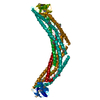
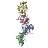
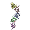
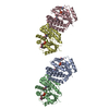
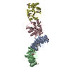
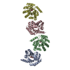
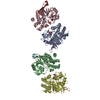
| Title | Crystal Structure of the BAR-PH domain of human APPL1 | ||||||
 Components Components | Adapter protein containing PH domain, PTB domain and leucine zipper motif 1 | ||||||
 Keywords Keywords | PROTEIN BINDING / APPL / BAR domain / PH domain | ||||||
| Function / homology |  Function and homology information Function and homology informationnegative regulation of Fc-gamma receptor signaling pathway involved in phagocytosis / positive regulation of macropinocytosis / adiponectin-activated signaling pathway / macropinosome / regulation of fibroblast migration / regulation of glucose import / maintenance of synapse structure / protein kinase B binding / signaling / positive regulation of melanin biosynthetic process ...negative regulation of Fc-gamma receptor signaling pathway involved in phagocytosis / positive regulation of macropinocytosis / adiponectin-activated signaling pathway / macropinosome / regulation of fibroblast migration / regulation of glucose import / maintenance of synapse structure / protein kinase B binding / signaling / positive regulation of melanin biosynthetic process / regulation of toll-like receptor 4 signaling pathway / vesicle membrane / positive regulation of cytokine production involved in inflammatory response / early phagosome / Caspase activation via Dependence Receptors in the absence of ligand / cellular response to hepatocyte growth factor stimulus / intracellular vesicle / phosphatidylserine binding / beta-tubulin binding / regulation of innate immune response / regulation of G1/S transition of mitotic cell cycle / regulation of protein localization to plasma membrane / ruffle / phosphatidylinositol binding / transforming growth factor beta receptor signaling pathway / positive regulation of glucose import / protein import into nucleus / presynapse / insulin receptor signaling pathway / cytoplasmic vesicle / early endosome membrane / postsynapse / early endosome / endosome membrane / endosome / cell cycle / glutamatergic synapse / protein-containing complex binding / signal transduction / protein homodimerization activity / extracellular exosome / membrane / identical protein binding / nucleus / plasma membrane / cytosol / cytoplasmSimilarity search - Function | ||||||
| Biological species |  Homo sapiens (human) Homo sapiens (human) | ||||||
| Method | X-RAY DIFFRACTION / SYNCHROTRON / SAD / Resolution: 2.6 Å | ||||||
 Authors Authors | Li, J. / Mao, X. / Dong, L.Q. / Liu, F. / Tong, L. | ||||||
 Citation Citation | Journal: Structure / Year: 2007 Title: Crystal Structures of the BAR-PH and PTB Domains of Human APPL1 Authors: Li, J. / Mao, X. / Dong, L.Q. / Liu, F. / Tong, L. | ||||||
| History |
|
- Structure visualization
| Structure viewer | Molecule: MolmilJmol/JSmol |
|---|
- Downloads & links
-Download
| PDBx/mmCIF format | 2elb.cif.gz | 80 KB | Display | PDBx/mmCIF format |
|---|---|---|---|---|
| PDB format | pdb2elb.ent.gz | 64.2 KB | Display | PDB format |
| PDBx/mmJSON format | 2elb.json.gz | Tree view | PDBx/mmJSON format | |
| Others |  Other downloads Other downloads |
-Validation report
| Arichive directory | https://data.pdbj.org/pub/pdb/validation_reports/el/2elbftp://data.pdbj.org/pub/pdb/validation_reports/el/2elb | HTTPS FTP |
|---|
-Related structure data


-Links
 PDBj
PDBj


- Assembly
| Deposited unit | 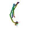
| ||||||||
|---|---|---|---|---|---|---|---|---|---|
| 1 | 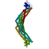
| ||||||||
| Unit cell |
| ||||||||
| Details | The biological assembly is a dimer. |
-Components
| #1: Protein | / APPL1 / Dip13 alpha / DCC-interacting protein 13 alpha Mass: 45987.117 Da / Num. of mol.: 1 / Fragment: The BAR-PH domain Source method: isolated from a genetically manipulated source Source: (gene. exp.) Homo sapiens (human) / Plasmid: pET28a / Species (production host): Escherichia coli / Production host:  Escherichia coli BL21(DE3) (bacteria) / Strain (production host): BL21(DE3) / References: UniProt: Q9UKG1 Escherichia coli BL21(DE3) (bacteria) / Strain (production host): BL21(DE3) / References: UniProt: Q9UKG1 |
|---|
-Experimental details
-Experiment
| Experiment | Method: X-RAY DIFFRACTION / Number of used crystals: 1 |
|---|
- Sample preparation
| Crystal | Density Matthews: 2.11 Å3/Da / Density % sol: 41.7 % |
|---|---|
| Crystal grow | Temperature: 293 K / Method: vapor diffusion, sitting drop / pH: 7.5 Details: 0.1M HEPES (pH 7.5), 22% (w/v) polyacrylic acid 5100, 20mM magnesium chloride, VAPOR DIFFUSION, SITTING DROP, temperature 293K |
-Data collection
| Diffraction | Mean temperature: 100 K |
|---|---|
| Diffraction source | Source: SYNCHROTRON / Site: NSLS  / Beamline: X4A / Wavelength: 0.97934 Å / Beamline: X4A / Wavelength: 0.97934 Å |
| Detector | Type: ADSC QUANTUM 4 / Detector: CCD / Details: mirrors |
| Radiation | Protocol: SINGLE WAVELENGTH / Monochromatic (M) / Laue (L): M / Scattering type: x-ray |
| Radiation wavelength | Wavelength: 0.97934 Å / Relative weight: 1 |
| Reflection | Resolution: 2.6→30 Å / Num. obs: 22741 / % possible obs: 98.3 % / Observed criterion σ(F): 2 / Observed criterion σ(I): 1 / Redundancy: 6.4 % / Biso Wilson estimate: 29.1 Å2 / Rmerge(I) obs: 0.067 / Net I/σ(I): 27.5853 |
| Reflection shell | Resolution: 2.6→2.69 Å / Redundancy: 6 % / Rmerge(I) obs: 0.339 / Mean I/σ(I) obs: 5.94 / Num. unique all: 2267 / % possible all: 98.4 |
- Processing
| Software |
| ||||||||||||||||||||||||||||||||||||||||||||||||||||||||||||||||||||||||||||||||
|---|---|---|---|---|---|---|---|---|---|---|---|---|---|---|---|---|---|---|---|---|---|---|---|---|---|---|---|---|---|---|---|---|---|---|---|---|---|---|---|---|---|---|---|---|---|---|---|---|---|---|---|---|---|---|---|---|---|---|---|---|---|---|---|---|---|---|---|---|---|---|---|---|---|---|---|---|---|---|---|---|---|
| Refinement | Method to determine structure: SAD / Resolution: 2.6→29.84 Å / Rfactor Rfree error: 0.009 / Data cutoff high absF: 594181.77 / Data cutoff low absF: 0 / Isotropic thermal model: RESTRAINED / Cross valid method: THROUGHOUT / σ(F): 1.5
| ||||||||||||||||||||||||||||||||||||||||||||||||||||||||||||||||||||||||||||||||
| Solvent computation | Solvent model: FLAT MODEL / Bsol: 22.6318 Å2 / ksol: 0.304871 e/Å3 | ||||||||||||||||||||||||||||||||||||||||||||||||||||||||||||||||||||||||||||||||
| Displacement parameters | Biso mean: 44.9 Å2
| ||||||||||||||||||||||||||||||||||||||||||||||||||||||||||||||||||||||||||||||||
| Refine analyze |
| ||||||||||||||||||||||||||||||||||||||||||||||||||||||||||||||||||||||||||||||||
| Refinement step | Cycle: LAST / Resolution: 2.6→29.84 Å
| ||||||||||||||||||||||||||||||||||||||||||||||||||||||||||||||||||||||||||||||||
| Refine LS restraints |
| ||||||||||||||||||||||||||||||||||||||||||||||||||||||||||||||||||||||||||||||||
| LS refinement shell | Resolution: 2.6→2.69 Å / Rfactor Rfree error: 0.032 / Total num. of bins used: 10
| ||||||||||||||||||||||||||||||||||||||||||||||||||||||||||||||||||||||||||||||||
| Xplor file |
|
