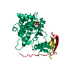
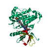
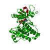
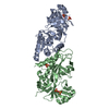
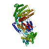
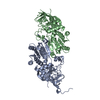
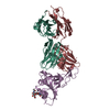
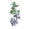
[English] 日本語
Yorodumi
- PDB-2eb2: Crystal structure of mutated EGFR kinase domain (G719S) -

+
Open data


ID or keywords:

Loading...

-
Basic information

Entry
Database: PDB / ID: 2eb2
TitleCrystal structure of mutated EGFR kinase domain (G719S)
ComponentsEpidermal growth factor receptor
KeywordsTRANSFERASE / Kinase / Tyrosine-protein kinase / Receptor / Phosphorylation / ATP-binding / Disease mutation / Transmembrane / Cell cycle / EC 2.7.10.1 / Structural Genomics / NPPSFA / National Project on Protein Structural and Functional Analyses / RIKEN Structural Genomics/Proteomics Initiative / RSGI
Function / homology
Function and homology information


response to hydroxyisoflavone / multivesicular body, internal vesicle lumen / positive regulation of prolactin secretion / negative regulation of cardiocyte differentiation / positive regulation of protein kinase C activity / diterpenoid metabolic process / Shc-EGFR complex / ovulation cycle / Inhibition of Signaling by Overexpressed EGFR / epidermal growth factor receptor activity ...response to hydroxyisoflavone / multivesicular body, internal vesicle lumen / positive regulation of prolactin secretion / negative regulation of cardiocyte differentiation / positive regulation of protein kinase C activity / diterpenoid metabolic process / Shc-EGFR complex / ovulation cycle / Inhibition of Signaling by Overexpressed EGFR / epidermal growth factor receptor activity / EGFR interacts with phospholipase C-gamma / positive regulation of mucus secretion / response to UV-A / epidermal growth factor binding / PLCG1 events in ERBB2 signaling / tongue development / midgut development / ERBB2-EGFR signaling pathway / hydrogen peroxide metabolic process / PTK6 promotes HIF1A stabilization / digestive tract morphogenesis / regulation of nitric-oxide synthase activity / morphogenesis of an epithelial fold / ERBB2 Activates PTK6 Signaling / intracellular vesicle / Signaling by EGFR / response to cobalamin / transmembrane receptor protein tyrosine kinase activator activity / protein tyrosine kinase activator activity / negative regulation of epidermal growth factor receptor signaling pathway / Signaling by ERBB4 / regulation of phosphatidylinositol 3-kinase/protein kinase B signal transduction / eyelid development in camera-type eye / protein insertion into membrane / cerebral cortex cell migration / ERBB2 Regulates Cell Motility / regulation of JNK cascade / : / PI3K events in ERBB2 signaling / positive regulation of cyclin-dependent protein serine/threonine kinase activity / negative regulation of mitotic cell cycle / hair follicle development / MAP kinase kinase kinase activity / Estrogen-dependent nuclear events downstream of ESR-membrane signaling / embryonic placenta development / positive regulation of bone resorption / positive regulation of G1/S transition of mitotic cell cycle / GAB1 signalosome / positive regulation of nitric oxide mediated signal transduction / salivary gland morphogenesis / peptidyl-tyrosine autophosphorylation / regulation of peptidyl-tyrosine phosphorylation / positive regulation of phosphorylation / positive regulation of glial cell proliferation / positive regulation of vasoconstriction / Signaling by ERBB2 / cellular response to epidermal growth factor stimulus / cellular response to cadmium ion / GRB2 events in EGFR signaling / SHC1 events in EGFR signaling / positive regulation of DNA repair / EGFR Transactivation by Gastrin / GRB2 events in ERBB2 signaling / TFAP2 (AP-2) family regulates transcription of growth factors and their receptors / transmembrane receptor protein tyrosine kinase activity / SHC1 events in ERBB2 signaling / ossification / positive regulation of synaptic transmission, glutamatergic / neurogenesis / cellular response to dexamethasone stimulus / basal plasma membrane / regulation of ERK1 and ERK2 cascade / neuron projection morphogenesis / positive regulation of superoxide anion generation / positive regulation of DNA replication / Signal transduction by L1 / epithelial cell proliferation / cellular response to estradiol stimulus / NOTCH3 Activation and Transmission of Signal to the Nucleus / positive regulation of epithelial cell proliferation / astrocyte activation / liver regeneration / positive regulation of protein localization to plasma membrane / EGFR downregulation / cell surface receptor protein tyrosine kinase signaling pathway / cellular response to amino acid stimulus / positive regulation of smooth muscle cell proliferation / Signaling by ERBB2 TMD/JMD mutants / positive regulation of MAP kinase activity / clathrin-coated endocytic vesicle membrane / lung development / Constitutive Signaling by EGFRvIII / Signaling by ERBB2 ECD mutants / epidermal growth factor receptor signaling pathway / Signaling by ERBB2 KD Mutants / negative regulation of protein catabolic process / receptor protein-tyrosine kinase / Downregulation of ERBB2 signaling / kinase binding / ruffle membrane
Similarity search - Function
: / Epidermal growth factor receptor transmembrane-juxtamembrane segment / Tyrosine protein kinase, EGF/ERB/XmrK receptor / Growth factor receptor domain 4 / Growth factor receptor domain IV / Receptor L-domain / Furin-like cysteine-rich domain / Receptor L-domain superfamily / Furin-like cysteine rich region / Receptor L domain ...: / Epidermal growth factor receptor transmembrane-juxtamembrane segment / Tyrosine protein kinase, EGF/ERB/XmrK receptor / Growth factor receptor domain 4 / Growth factor receptor domain IV / Receptor L-domain / Furin-like cysteine-rich domain / Receptor L-domain superfamily / Furin-like cysteine rich region / Receptor L domain / Furin-like repeat / Furin-like repeats / Growth factor receptor cysteine-rich domain superfamily / Tyrosine-protein kinase, catalytic domain / Tyrosine kinase, catalytic domain / Tyrosine protein kinases specific active-site signature. / Tyrosine-protein kinase, active site / Protein tyrosine and serine/threonine kinase / Serine-threonine/tyrosine-protein kinase, catalytic domain / Transferase(Phosphotransferase) domain 1 / Transferase(Phosphotransferase); domain 1 / Phosphorylase Kinase; domain 1 / Phosphorylase Kinase; domain 1 / Protein kinase, ATP binding site / Protein kinases ATP-binding region signature. / Protein kinase domain profile. / Protein kinase domain / Protein kinase-like domain superfamily / 2-Layer Sandwich / Orthogonal Bundle / Mainly Alpha / Alpha Beta
Similarity search - Domain/homology
Epidermal growth factor receptor
Similarity search - Component
Biological speciesHomo sapiens (human)
MethodX-RAY DIFFRACTION / SYNCHROTRON / MOLECULAR REPLACEMENT / Resolution: 2.5 Å
AuthorsYoshikawa, S. / Kukimoto-Niino, M. / Chen, L. / Liu, Z.J. / Wang, B.C. / Shirouzu, M. / Senba, K. / Yamamoto, T. / Yokoyama, S. / RIKEN Structural Genomics/Proteomics Initiative (RSGI)
CitationJournal: Oncogene / Year: 2012
Title: Structural basis for the altered drug sensitivities of non-small cell lung cancer-associated mutants of human epidermal growth factor receptor
Authors: Yoshikawa, S. / Kukimoto-Niino, M. / Parker, L. / Handa, N. / Terada, T. / Fujimoto, T. / Terazawa, Y. / Wakiyama, M. / Sato, M. / Sano, S. / Kobayashi, T. / Tanaka, T. / Chen, L. / Liu, Z.J. ...Authors: Yoshikawa, S. / Kukimoto-Niino, M. / Parker, L. / Handa, N. / Terada, T. / Fujimoto, T. / Terazawa, Y. / Wakiyama, M. / Sato, M. / Sano, S. / Kobayashi, T. / Tanaka, T. / Chen, L. / Liu, Z.J. / Wang, B.C. / Shirouzu, M. / Kawa, S. / Semba, K. / Yamamoto, T. / Yokoyama, S.
History
DepositionFeb 6, 2007Deposition site: PDBJ / Processing site: PDBJ
Revision 1.0Feb 12, 2008Provider: repository / Type: Initial release
Revision 1.1Jul 13, 2011Group: Version format compliance
Revision 1.2Mar 7, 2012Group: Database references
Revision 1.3Oct 25, 2023Group: Data collection / Database references / Refinement description
Category: chem_comp_atom / chem_comp_bond ...chem_comp_atom / chem_comp_bond / database_2 / pdbx_initial_refinement_model / struct_ref_seq_dif
Item: _database_2.pdbx_DOI / _database_2.pdbx_database_accession / _struct_ref_seq_dif.details

-
Structure visualization

Structure viewerMolecule:
MolmilJmol/JSmol

Downloads & links

-
Assembly

Deposited unit
A: Epidermal growth factor receptor


Theoretical massNumber of molelcules
Total (without water)38,0081
Polymers38,0081
Non-polymers00
Water28816
1
A: Epidermal growth factor receptor

A: Epidermal growth factor receptor


Theoretical massNumber of molelcules
Total (without water)76,0162
Polymers76,0162
Non-polymers00
Water362
TypeNameSymmetry operationNumber
identity operation1_555x,y,z1
crystal symmetry operation20_555-z+1/2,x+1/2,-y+1/21
Unit cell
Length a, b, c (Å)145.470, 145.470, 145.470
Angle α, β, γ (deg.)90.00, 90.00, 90.00
Int Tables number197
Space group name H-MI23

-
Components

#1: Protein Epidermal growth factor receptor / / Receptor tyrosine-protein kinase ErbB-1


Mass: 38007.953 Da / Num. of mol.: 1 / Fragment: kinase domain, UNP residues 695-1022 / Mutation: G719S
Source method: isolated from a genetically manipulated source
Source: (gene. exp.) Homo sapiens (human) / Gene: EGFR / Production host: Spodoptera frugiperda (fall armyworm) / Strain (production host): Sf9
References: UniProt: P00533, receptor protein-tyrosine kinase
#2: Water ChemComp-HOH / water / Water


Mass: 18.015 Da / Num. of mol.: 16 / Source method: isolated from a natural source / Formula: H2O
Sequence detailsTHE RESIDUE IS SOMATIC MUTATION IN LUNG CANCER.

-
Experimental details

-
Experiment

ExperimentMethod: X-RAY DIFFRACTION / Number of used crystals: 1

-
Sample preparation

CrystalDensity Matthews: 3.37 Å3/Da / Density % sol: 63.53 %
Crystal growTemperature: 293 K / Method: vapor diffusion, hanging drop / pH: 7
Details: 0.1M MES, 1.1M Na/K tartrate, pH 7.0, VAPOR DIFFUSION, HANGING DROP, temperature 293K

-
Data collection

DiffractionMean temperature: 100 K
Diffraction sourceSource: SYNCHROTRON / Site: APS / Beamline: 22-ID / Wavelength: 0.97243 Å
DetectorType: MARRESEARCH / Detector: CCD / Date: Aug 6, 2006
RadiationMonochromator: Si double crystal / Protocol: SINGLE WAVELENGTH / Monochromatic (M) / Laue (L): M / Scattering type: x-ray
Radiation wavelengthWavelength: 0.97243 Å / Relative weight: 1
ReflectionResolution: 2.5→50 Å / Num. obs: 17866 / % possible obs: 100 % / Observed criterion σ(I): -3 / Redundancy: 43.094 % / Biso Wilson estimate: 46 Å2 / Rsym value: 0.137 / Net I/σ(I): 35.357
Reflection shellResolution: 2.5→2.59 Å / Redundancy: 33.3 % / Mean I/σ(I) obs: 9.75 / Rsym value: 0.484 / % possible all: 100

-
Processing

Software
NameVersionClassification
CNS1.1refinement
HKL-2000data collection
DENZOdata reduction
SCALEPACKdata scaling
MOLREPphasing
RefinementMethod to determine structure: MOLECULAR REPLACEMENT
Starting model: PDB ENTRY 1M14
Resolution: 2.5→46 Å / Rfactor Rfree error: 0.006 / Data cutoff high absF: 2990657.37 / Data cutoff low absF: 0 / Isotropic thermal model: RESTRAINED / Cross valid method: THROUGHOUT / σ(F): 0 / Stereochemistry target values: Engh & Huber
RfactorNum. reflection% reflectionSelection details
Rfree0.253 1755 9.8 %RANDOM
Rwork0.198 ---
obs0.198 17863 99.8 %-
Solvent computationSolvent model: FLAT MODEL / Bsol: 48.7334 Å2 / ksol: 0.367323 e/Å3
Displacement parametersBiso mean: 58.2 Å2
Baniso -1Baniso -2Baniso -3
1-0 Å20 Å20 Å2
2--0 Å20 Å2
3---0 Å2
Refine analyze
FreeObs
Luzzati coordinate error0.38 Å0.28 Å
Luzzati d res low-5 Å
Luzzati sigma a0.32 Å0.22 Å
Refinement stepCycle: LAST / Resolution: 2.5→46 Å
ProteinNucleic acidLigandSolventTotal
Num. atoms2443 0 0 16 2459
Refine LS restraints
Refine-IDTypeDev idealDev ideal target
X-RAY DIFFRACTIONc_bond_d0.01
X-RAY DIFFRACTIONc_bond_d_na
X-RAY DIFFRACTIONc_bond_d_prot
X-RAY DIFFRACTIONc_angle_d
X-RAY DIFFRACTIONc_angle_d_na
X-RAY DIFFRACTIONc_angle_d_prot
X-RAY DIFFRACTIONc_angle_deg1.5
X-RAY DIFFRACTIONc_angle_deg_na
X-RAY DIFFRACTIONc_angle_deg_prot
X-RAY DIFFRACTIONc_dihedral_angle_d23.6
X-RAY DIFFRACTIONc_dihedral_angle_d_na
X-RAY DIFFRACTIONc_dihedral_angle_d_prot
X-RAY DIFFRACTIONc_improper_angle_d1.07
X-RAY DIFFRACTIONc_improper_angle_d_na
X-RAY DIFFRACTIONc_improper_angle_d_prot
X-RAY DIFFRACTIONc_mcbond_it6.741.5
X-RAY DIFFRACTIONc_mcangle_it8.832
X-RAY DIFFRACTIONc_scbond_it10.492
X-RAY DIFFRACTIONc_scangle_it12.332.5
LS refinement shellResolution: 2.5→2.66 Å / Rfactor Rfree error: 0.019 / Total num. of bins used: 6
RfactorNum. reflection% reflection
Rfree0.33 315 10.8 %
Rwork0.254 2613 -
obs--100 %
Xplor file
Refine-IDSerial noParam fileTopol file
X-RAY DIFFRACTION1protein_rep.paramprotein.top
X-RAY DIFFRACTION2water_rep.paramwater.top

+
About Yorodumi

-
News

-
Feb 9, 2022. New format data for meta-information of EMDB entries

New format data for meta-information of EMDB entries

  • Version 3 of the EMDB header file is now the official format.
  • The previous official version 1.9 will be removed from the archive.

Related info.:EMDB header

External links:wwPDB to switch to version 3 of the EMDB data model

-
Aug 12, 2020. Covid-19 info

Covid-19 info

URL: https://pdbj.org/emnavi/covid19.php

New page: Covid-19 featured information page in EM Navigator.

Related info.:Covid-19 info / Mar 5, 2020. Novel coronavirus structure data

+
Mar 5, 2020. Novel coronavirus structure data

Novel coronavirus structure data

Related info.:Yorodumi Speices / Aug 12, 2020. Covid-19 info

External links:COVID-19 featured content - PDBj / Molecule of the Month (242):Coronavirus Proteases

+
Jan 31, 2019. EMDB accession codes are about to change! (news from PDBe EMDB page)

EMDB accession codes are about to change! (news from PDBe EMDB page)

  • The allocation of 4 digits for EMDB accession codes will soon come to an end. Whilst these codes will remain in use, new EMDB accession codes will include an additional digit and will expand incrementally as the available range of codes is exhausted. The current 4-digit format prefixed with “EMD-” (i.e. EMD-XXXX) will advance to a 5-digit format (i.e. EMD-XXXXX), and so on. It is currently estimated that the 4-digit codes will be depleted around Spring 2019, at which point the 5-digit format will come into force.
  • The EM Navigator/Yorodumi systems omit the EMD- prefix.

Related info.:Q: What is EMD? / ID/Accession-code notation in Yorodumi/EM Navigator

External links:EMDB Accession Codes are Changing Soon! / Contact to PDBj

+
Jul 12, 2017. Major update of PDB

Major update of PDB

  • wwPDB released updated PDB data conforming to the new PDBx/mmCIF dictionary.
  • This is a major update changing the version number from 4 to 5, and with Remediation, in which all the entries are updated.
  • In this update, many items about electron microscopy experimental information are reorganized (e.g. em_software).
  • Now, EM Navigator and Yorodumi are based on the updated data.

External links:wwPDB Remediation / Enriched Model Files Conforming to OneDep Data Standards Now Available in the PDB FTP Archive

-
Yorodumi

Thousand views of thousand structures

  • Yorodumi is a browser for structure data from EMDB, PDB, SASBDB, etc.
  • This page is also the successor to EM Navigator detail page, and also detail information page/front-end page for Omokage search.
  • The word "yorodu" (or yorozu) is an old Japanese word meaning "ten thousand". "mi" (miru) is to see.

Related info.:EMDB / PDB / SASBDB / Comparison of 3 databanks / Yorodumi Search / Aug 31, 2016. New EM Navigator & Yorodumi / Yorodumi Papers / Jmol/JSmol / Function and homology information / Changes in new EM Navigator and Yorodumi

Read more