 Movie
Movie Controller

[English] 日本語
 Yorodumi
- PDB-2e4z: Crystal structure of the ligand-binding region of the group III m... -
+ Open data

- Basic information
| Entry | Database: PDB / ID: 2e4z | ||||||
|---|---|---|---|---|---|---|---|
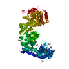
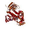
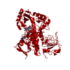
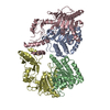
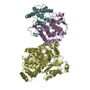
| Title | Crystal structure of the ligand-binding region of the group III metabotropic glutamate receptor | ||||||
 Components Components | Metabotropic glutamate receptor 7 | ||||||
 Keywords Keywords | SIGNALING PROTEIN / G-protein-coupled receptor / neuron / central nerve system | ||||||
| Function / homology |  Function and homology information Function and homology informationgroup III metabotropic glutamate receptor activity / : / Class C/3 (Metabotropic glutamate/pheromone receptors) / adenylate cyclase-inhibiting G protein-coupled glutamate receptor signaling pathway / adenylate cyclase inhibitor activity / glycosylation / : / negative regulation of glutamate secretion / adenylate cyclase inhibiting G protein-coupled glutamate receptor activity / serine binding ...group III metabotropic glutamate receptor activity / : / Class C/3 (Metabotropic glutamate/pheromone receptors) / adenylate cyclase-inhibiting G protein-coupled glutamate receptor signaling pathway / adenylate cyclase inhibitor activity / glycosylation / : / negative regulation of glutamate secretion / adenylate cyclase inhibiting G protein-coupled glutamate receptor activity / serine binding / conditioned taste aversion / short-term memory / G protein-coupled glutamate receptor signaling pathway / nervous system process / glutamate receptor activity / G alpha (i) signalling events / glutamate binding / regulation of synaptic vesicle exocytosis / adult behavior / axon development / transmission of nerve impulse / presynaptic active zone / plasma membrane => GO:0005886 / associative learning / calcium channel regulator activity / asymmetric synapse / GABA-ergic synapse / voltage-gated calcium channel activity / behavioral fear response / multicellular organismal response to stress / axon terminus / rough endoplasmic reticulum / regulation of synaptic transmission, glutamatergic / dendritic shaft / PDZ domain binding / sensory perception of sound / terminal bouton / memory / calcium-dependent protein binding / presynaptic membrane / cell cortex / chemical synaptic transmission / postsynaptic membrane / negative regulation of neuron apoptotic process / membrane => GO:0016020 / learning or memory / receptor complex / protein dimerization activity / calmodulin binding / axon / neuronal cell body / glutamatergic synapse / synapse / dendrite / Golgi apparatus / cell surface / protein-containing complex / membrane / identical protein binding / plasma membraneSimilarity search - Function | ||||||
| Biological species |  Rattus norvegicus (Norway rat) Rattus norvegicus (Norway rat) | ||||||
| Method | X-RAY DIFFRACTION / SYNCHROTRON / MOLECULAR REPLACEMENT / Resolution: 3.3 Å | ||||||
 Authors Authors | Muto, T. / Tsuchiya, D. / Morikawa, K. / Jingami, H. | ||||||
 Citation Citation | Journal: Proc.Natl.Acad.Sci.Usa / Year: 2007 Title: Structures of the extracellular regions of the group II/III metabotropic glutamate receptors Authors: Muto, T. / Tsuchiya, D. / Morikawa, K. / Jingami, H. | ||||||
| History |
|
- Structure visualization
| Structure viewer | Molecule: MolmilJmol/JSmol |
|---|
- Downloads & links
-Download
| PDBx/mmCIF format | 2e4z.cif.gz | 98.5 KB | Display | PDBx/mmCIF format |
|---|---|---|---|---|
| PDB format | pdb2e4z.ent.gz | 72.6 KB | Display | PDB format |
| PDBx/mmJSON format | 2e4z.json.gz | Tree view | PDBx/mmJSON format | |
| Others |  Other downloads Other downloads |
-Validation report
| Arichive directory | https://data.pdbj.org/pub/pdb/validation_reports/e4/2e4zftp://data.pdbj.org/pub/pdb/validation_reports/e4/2e4z | HTTPS FTP |
|---|
-Related structure data
| Related structure data | 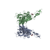 2e4uC 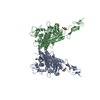 2e4vC 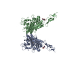 2e4wC 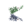 2e4xC 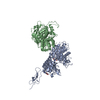 2e4yC  1ewkS C: citing same article ( S: Starting model for refinement |
|---|---|
| Similar structure data |


-Links
 PDBj
PDBj


- Assembly
| Deposited unit | 
| ||||||||
|---|---|---|---|---|---|---|---|---|---|
| 1 | 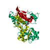
| ||||||||
| Unit cell |
| ||||||||
| Details | The second part of the biological assembly is generated by the two fold axis: X-Y,-Y,-1/3-Z |
-Components
| #1: Protein | / mGluR7 / group III metabotropic glutamate receptor subtype 7 Mass: 56030.871 Da / Num. of mol.: 1 / Fragment: ligand-binding region, residues 33-521 Source method: isolated from a genetically manipulated source Source: (gene. exp.) Rattus norvegicus (Norway rat) / Cell line (production host): HIGH FIVE / Production host:  Trichoplusia ni (cabbage looper) / References: UniProt: P35400 Trichoplusia ni (cabbage looper) / References: UniProt: P35400 |
|---|---|
| #2: Chemical | ChemComp-MES / MES (buffer)  Mass: 195.237 Da / Num. of mol.: 1 / Source method: obtained synthetically / Formula: C6H13NO4S / Comment: pH buffer*YM Mass: 195.237 Da / Num. of mol.: 1 / Source method: obtained synthetically / Formula: C6H13NO4S / Comment: pH buffer*YM |
-Experimental details
-Experiment
| Experiment | Method: X-RAY DIFFRACTION / Number of used crystals: 1 |
|---|
- Sample preparation
| Crystal | Density Matthews: 2.52 Å3/Da / Density % sol: 51.1 % |
|---|---|
| Crystal grow | Temperature: 298 K / Method: vapor diffusion, hanging drop / pH: 5.8 Details: 0.02M Citrate, 0.08M MES, 0.01M Glutamate, 2.2M ammonium sulfate, pH 5.8, VAPOR DIFFUSION, HANGING DROP, temperature 298K |
-Data collection
| Diffraction | Mean temperature: 100 K |
|---|---|
| Diffraction source | Source: SYNCHROTRON / Site: SPring-8  / Beamline: BL41XU / Wavelength: 1 Å / Beamline: BL41XU / Wavelength: 1 Å |
| Detector | Type: ADSC QUANTUM 315 / Detector: CCD / Date: Dec 1, 2004 |
| Radiation | Monochromator: SI / Protocol: SINGLE WAVELENGTH / Monochromatic (M) / Laue (L): M / Scattering type: x-ray |
| Radiation wavelength | Wavelength: 1 Å / Relative weight: 1 |
| Reflection | Resolution: 3.3→100 Å / Num. all: 8887 / Num. obs: 8887 / % possible obs: 100 % / Observed criterion σ(F): 0 / Observed criterion σ(I): 0 / Rmerge(I) obs: 0.112 / Rsym value: 0.106 / Net I/σ(I): 5.4 |
| Reflection shell | Resolution: 3.3→3.48 Å / Rmerge(I) obs: 0.54 / Mean I/σ(I) obs: 1.4 / % possible all: 100 |
- Processing
| Software |
| ||||||||||||||||||||||||||||||||||||||||||||||||||||||||||||||||||||||||||||||||||||||||||||||||||||||||||||||||||||||||||||||||||||||||||||||||||||||||||||||||||||||||||
|---|---|---|---|---|---|---|---|---|---|---|---|---|---|---|---|---|---|---|---|---|---|---|---|---|---|---|---|---|---|---|---|---|---|---|---|---|---|---|---|---|---|---|---|---|---|---|---|---|---|---|---|---|---|---|---|---|---|---|---|---|---|---|---|---|---|---|---|---|---|---|---|---|---|---|---|---|---|---|---|---|---|---|---|---|---|---|---|---|---|---|---|---|---|---|---|---|---|---|---|---|---|---|---|---|---|---|---|---|---|---|---|---|---|---|---|---|---|---|---|---|---|---|---|---|---|---|---|---|---|---|---|---|---|---|---|---|---|---|---|---|---|---|---|---|---|---|---|---|---|---|---|---|---|---|---|---|---|---|---|---|---|---|---|---|---|---|---|---|---|---|---|
| Refinement | Method to determine structure: MOLECULAR REPLACEMENT Starting model: PDB ENTRY 1EWK Resolution: 3.3→12 Å / Cor.coef. Fo:Fc: 0.882 / Cor.coef. Fo:Fc free: 0.823 / SU B: 33.152 / SU ML: 0.579 / TLS residual ADP flag: LIKELY RESIDUAL / Cross valid method: THROUGHOUT / σ(F): 3 / σ(I): 3 / ESU R Free: 0.702 / Stereochemistry target values: Engh & Huber
| ||||||||||||||||||||||||||||||||||||||||||||||||||||||||||||||||||||||||||||||||||||||||||||||||||||||||||||||||||||||||||||||||||||||||||||||||||||||||||||||||||||||||||
| Solvent computation | Ion probe radii: 0.8 Å / Shrinkage radii: 0.8 Å / VDW probe radii: 1.4 Å / Solvent model: MASK | ||||||||||||||||||||||||||||||||||||||||||||||||||||||||||||||||||||||||||||||||||||||||||||||||||||||||||||||||||||||||||||||||||||||||||||||||||||||||||||||||||||||||||
| Displacement parameters | Biso mean: 41.703 Å2
| ||||||||||||||||||||||||||||||||||||||||||||||||||||||||||||||||||||||||||||||||||||||||||||||||||||||||||||||||||||||||||||||||||||||||||||||||||||||||||||||||||||||||||
| Refinement step | Cycle: LAST / Resolution: 3.3→12 Å
| ||||||||||||||||||||||||||||||||||||||||||||||||||||||||||||||||||||||||||||||||||||||||||||||||||||||||||||||||||||||||||||||||||||||||||||||||||||||||||||||||||||||||||
| Refine LS restraints |
| ||||||||||||||||||||||||||||||||||||||||||||||||||||||||||||||||||||||||||||||||||||||||||||||||||||||||||||||||||||||||||||||||||||||||||||||||||||||||||||||||||||||||||
| LS refinement shell | Resolution: 3.3→3.379 Å / Total num. of bins used: 20 /
| ||||||||||||||||||||||||||||||||||||||||||||||||||||||||||||||||||||||||||||||||||||||||||||||||||||||||||||||||||||||||||||||||||||||||||||||||||||||||||||||||||||||||||
| Refinement TLS params. | Method: refined / Refine-ID: X-RAY DIFFRACTION
| ||||||||||||||||||||||||||||||||||||||||||||||||||||||||||||||||||||||||||||||||||||||||||||||||||||||||||||||||||||||||||||||||||||||||||||||||||||||||||||||||||||||||||
| Refinement TLS group |
|
