 Movie
Movie Controller

[English] 日本語
 Yorodumi
- PDB-2dyq: Crystal Structure of the C-terminal Phophotyrosine Interaction Do... -
+ Open data

- Basic information
| Entry | Database: PDB / ID: 2dyq | ||||||
|---|---|---|---|---|---|---|---|
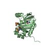
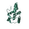
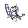
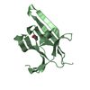
| Title | Crystal Structure of the C-terminal Phophotyrosine Interaction Domain of Human APBB3 | ||||||
 Components Components | Amyloid beta A4 precursor protein-binding family B member 3 | ||||||
 Keywords Keywords |  PROTEIN BINDING / Phosphotyrosine-interaction domain (PTB/PID) / Alzheimer's disease / Structural Genomics / NPPSFA / National Project on Protein Structural and Functional Analyses / RIKEN Structural Genomics/Proteomics Initiative / RSGI PROTEIN BINDING / Phosphotyrosine-interaction domain (PTB/PID) / Alzheimer's disease / Structural Genomics / NPPSFA / National Project on Protein Structural and Functional Analyses / RIKEN Structural Genomics/Proteomics Initiative / RSGI | ||||||
| Function / homology |  Function and homology information Function and homology informationlow-density lipoprotein particle receptor binding / positive regulation of protein secretion / actin cytoskeleton / amyloid-beta binding / nuclear body / regulation of DNA-templated transcription / membrane / nucleus / cytosol / cytoplasmSimilarity search - Function | ||||||
| Biological species |  Homo sapiens (human) Homo sapiens (human) | ||||||
| Method | X-RAY DIFFRACTION / SYNCHROTRON / SAD / Resolution: 3.1 Å | ||||||
 Authors Authors | Nishino, A. / Saijo, S. / Kishishita, S. / Shirouzu, M. / Yokoyama, S. / RIKEN Structural Genomics/Proteomics Initiative (RSGI) | ||||||
 Citation Citation | Journal: To be Published Title: Crystal Structure of the C-terminal Phophotyrosine Interaction Domain of Human APBB3 Authors: Saijo, S. / Nishino, A. / Kishishita, S. / Shirouzu, M. / Yokoyama, S. | ||||||
| History |
|
- Structure visualization
| Structure viewer | Molecule: MolmilJmol/JSmol |
|---|
- Downloads & links
-Download
| PDBx/mmCIF format | 2dyq.cif.gz | 32.1 KB | Display | PDBx/mmCIF format |
|---|---|---|---|---|
| PDB format | pdb2dyq.ent.gz | 24.6 KB | Display | PDB format |
| PDBx/mmJSON format | 2dyq.json.gz | Tree view | PDBx/mmJSON format | |
| Others |  Other downloads Other downloads |
-Validation report
| Arichive directory | https://data.pdbj.org/pub/pdb/validation_reports/dy/2dyqftp://data.pdbj.org/pub/pdb/validation_reports/dy/2dyq | HTTPS FTP |
|---|
-Related structure data
| Similar structure data | |
|---|---|
| Other databases |
-Links
 PDBj
PDBj

- Assembly
| Deposited unit | 
| ||||||||
|---|---|---|---|---|---|---|---|---|---|
| 1 |
| ||||||||
| Unit cell |
|
-Components
| #1: Protein | Mass: 15584.754 Da / Num. of mol.: 1 Fragment: C-terminal Phosphotyrosine interaction domain (PTB/PID) Source method: isolated from a genetically manipulated source Source: (gene. exp.) Homo sapiens (human) / Description: Cell-free protein synthesis / Gene: APBB3, FE65L2 / Plasmid: PK060110-10 / References: UniProt: O95704 |
|---|
-Experimental details
-Experiment
| Experiment | Method: X-RAY DIFFRACTION / Number of used crystals: 1 |
|---|
- Sample preparation
| Crystal | Density Matthews: 3.29 Å3/Da / Density % sol: 62.58 % |
|---|---|
| Crystal grow | Temperature: 293 K / Method: vapor diffusion, sitting drop / pH: 9 Details: 25% PEG 3350, 0.2M Ammonium Acetate, 0.1M Tris-HCl, pH 9.0, VAPOR DIFFUSION, SITTING DROP, temperature 293K |
-Data collection
| Diffraction | Mean temperature: 100 K |
|---|---|
| Diffraction source | Source: SYNCHROTRON / Site: SPring-8  / Beamline: BL26B2 / Wavelength: 0.97895 Å / Beamline: BL26B2 / Wavelength: 0.97895 Å |
| Detector | Type: RIGAKU JUPITER 210 / Detector: CCD / Date: Apr 19, 2006 / Details: mirrors |
| Radiation | Monochromator: Fixed exit Si double crystal monochromator / Protocol: SINGLE WAVELENGTH / Monochromatic (M) / Laue (L): M / Scattering type: x-ray |
| Radiation wavelength | Wavelength: 0.97895 Å / Relative weight: 1 |
| Reflection | Resolution: 3.1→71.25 Å / Num. obs: 4171 / % possible obs: 99.9 % / Observed criterion σ(I): 0 / Redundancy: 18.4 % / Biso Wilson estimate: 45.2 Å2 / Rsym value: 0.08 / Net I/σ(I): 51.3 |
| Reflection shell | Resolution: 3.1→3.15 Å / Redundancy: 20.2 % / Mean I/σ(I) obs: 8.6 / Num. unique all: 202 / Rsym value: 0.407 / % possible all: 100 |
- Processing
| Software |
| ||||||||||||||||||||||||||||||||||||||||||||||||||||||||||||||||||||||||||||||||||||||||||||||||||||||||||||||||||||||||||||||||||||||||||||||||||||||||||||||||||||||||||
|---|---|---|---|---|---|---|---|---|---|---|---|---|---|---|---|---|---|---|---|---|---|---|---|---|---|---|---|---|---|---|---|---|---|---|---|---|---|---|---|---|---|---|---|---|---|---|---|---|---|---|---|---|---|---|---|---|---|---|---|---|---|---|---|---|---|---|---|---|---|---|---|---|---|---|---|---|---|---|---|---|---|---|---|---|---|---|---|---|---|---|---|---|---|---|---|---|---|---|---|---|---|---|---|---|---|---|---|---|---|---|---|---|---|---|---|---|---|---|---|---|---|---|---|---|---|---|---|---|---|---|---|---|---|---|---|---|---|---|---|---|---|---|---|---|---|---|---|---|---|---|---|---|---|---|---|---|---|---|---|---|---|---|---|---|---|---|---|---|---|---|---|
| Refinement | Method to determine structure: SAD / Resolution: 3.1→50 Å / Cor.coef. Fo:Fc: 0.901 / Cor.coef. Fo:Fc free: 0.852 / SU B: 50.292 / SU ML: 0.387 / TLS residual ADP flag: LIKELY RESIDUAL / Isotropic thermal model: Isotropic / Cross valid method: THROUGHOUT / σ(F): 0 / ESU R: 1.37 / ESU R Free: 0.452 / Stereochemistry target values: MAXIMUM LIKELIHOOD
| ||||||||||||||||||||||||||||||||||||||||||||||||||||||||||||||||||||||||||||||||||||||||||||||||||||||||||||||||||||||||||||||||||||||||||||||||||||||||||||||||||||||||||
| Solvent computation | Ion probe radii: 0.8 Å / Shrinkage radii: 0.8 Å / VDW probe radii: 1.2 Å / Solvent model: MASK | ||||||||||||||||||||||||||||||||||||||||||||||||||||||||||||||||||||||||||||||||||||||||||||||||||||||||||||||||||||||||||||||||||||||||||||||||||||||||||||||||||||||||||
| Displacement parameters | Biso mean: 46.3 Å2
| ||||||||||||||||||||||||||||||||||||||||||||||||||||||||||||||||||||||||||||||||||||||||||||||||||||||||||||||||||||||||||||||||||||||||||||||||||||||||||||||||||||||||||
| Refinement step | Cycle: LAST / Resolution: 3.1→50 Å
| ||||||||||||||||||||||||||||||||||||||||||||||||||||||||||||||||||||||||||||||||||||||||||||||||||||||||||||||||||||||||||||||||||||||||||||||||||||||||||||||||||||||||||
| Refine LS restraints |
| ||||||||||||||||||||||||||||||||||||||||||||||||||||||||||||||||||||||||||||||||||||||||||||||||||||||||||||||||||||||||||||||||||||||||||||||||||||||||||||||||||||||||||
| LS refinement shell | Resolution: 3.105→3.185 Å / Total num. of bins used: 20
| ||||||||||||||||||||||||||||||||||||||||||||||||||||||||||||||||||||||||||||||||||||||||||||||||||||||||||||||||||||||||||||||||||||||||||||||||||||||||||||||||||||||||||
| Refinement TLS params. | Method: refined / Origin x: 32.012 Å / Origin y: 36.536 Å / Origin z: 11.352 Å
|
